In order to successfully approach any surgical reconstruction, whether it be for a wound, deficit, or condition originating from burns, trauma, cancer, infections, or congenital conditions, it is beneficial to think through the available reconstructive options using as has been discussed in "Thinking Like a Reconstructive Surgeon: The Reconstructive Ladder." This section will build upon this concept by going into greater detail about the relevant anatomy for flaps as well as the classification systems used to describe and organize them.
Relevant Anatomy[edit | edit source]
In order to understand the various flap classification systems, it is important to know the relevant anatomy. In particular, the skin vasculature, perforator type, and blood supply all play roles in appreciating how and why flaps are classified.
Skin Anatomy[edit | edit source]
Vasculature[edit | edit source]
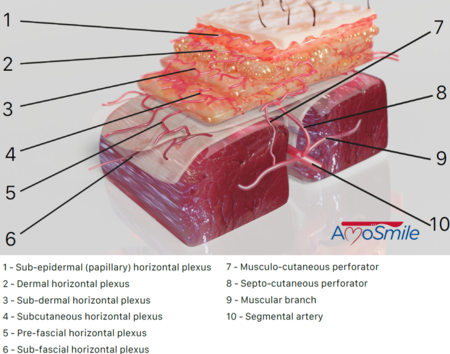
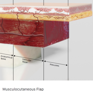
The skin receives its blood supply from a complex network of plexi and perforators that are perfused by larger underlying blood vessels. The figure below defines the various vascular networks and their relation to one another. It is important to note the sub-dermal plexus which is the primary source of blood supply to Random Pattern Flaps (see discussion on Random blood supply below). The larger perforators (i.e. musculocutaneous, septocutaneous), muscular branches, and segmental arteries create the axial blood supplies which feed into well-defined Axial Pattern flaps (see discussion on Axial blood supply below).
Perforator Anatomy[edit | edit source]
Perforators are small blood vessels that branch off source vessels and communicate with the various plexi of the fascia, subdermis, and dermis. These perforators supply specific units of skin and subcutaneous tissue known as "perforasomes" and knowledge of their distribution can be used to help design well-perfused flaps. The perforator type (see below for details) is an anatomically based description of the pathway that the perforating vessel travels in relation to surrounding anatomy and is important in guiding flap dissection. Both muscle/musculocutaneous and fascia/fasciocutaneous flaps have perforator types but they are not mutually exclusive.
- Direct Cutaneous: Cutaneous (direct) perforators course directly to the skin from the source or axial vessel and do not perforate through muscle or septi.
- Septocutaneous: Septocutaneous perforators course in between muscles via the intermuscular septi prior to reaching the skin.
- Musculocutaneous: Musculocutaneous perforators course through muscle prior to reaching the skin.
- Endosteal: These perforators arise from the intramedullary blood vessels within bone that then perforate the osseous cortex and go on to perfuse surrounding musculature and skin.
- Periosteal: These perforators arise from the periosteal blood vessels that surround the bone cortex which then course superficially to perfuse surrounding musculature and skin.
Blood Supply: Random vs. Axial[edit | edit source]
There are two primary types of bloody supply to flaps - Random and Axial.
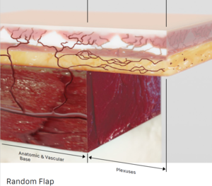
- Random: This type of flap is purely supplied by the subdermal plexus and other dermal plexi of the skin without a defined "axial" or "source" vessel that can be dissected out or isolated. Because of the "random" nature of the blood supply, these flaps are called Random Pattern Flaps.
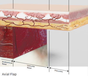
- Axial: This type of flap receives perfusion from a defined "source" vessel that is anatomically preserved and may be dissected out either for a pedicled or free flap. The term Axial is derived from the fact that there is an arterial "axis" through which the flap is perfused.
Common Flap Classification Systems[edit | edit source]
Mathes & Nahai (muscle/musculocutaneous)[edit | edit source]
- The Mathes & Nahai Muscle & Musculocutaneous Flap classification system is a commonly applied method of organizing and understanding the anatomy for various muscle-containing flaps. Understanding this classification system is not only important for being able to communicate about these types of flaps but also for appreciating the anatomical knowledge necessary to perform the flap dissections, elevations, and application. The system divides muscle and musculocutaneous flaps into 5 categories is based on the number and size of blood vessels that perfuse each flap. Below there are links to tables containing region-specific flaps and their classification with this system where appropriate.
- I: one Major blood supply
- II: one Major & one Minor blood supply
- III: two Major blood supplies
- IV: Segmental blood supply
- V: One Major & Segmental blood supply
Mathes & Nahai (fascia/fasciocutaneous)[edit | edit source]
- The Mathes & Nahai Fascia & Fasciocutaneous Flap classification system is a commonly applied method of organizing and understanding the anatomy for various fascia flaps. Understanding this classification system is not only important for being able to communicate about these types of flaps but also for appreciating the anatomical knowledge necessary to perform the flap dissections, elevations, and application. The system divides fascia and fasciocutaneous flaps into 3 categories based on the path that blood vessels travel on the way to perfusing the flap. Below there are links to tables containing region-specific flaps and their classification with this system where appropriate.
- A: Cutaneous (direct) perforator that travels directly to the skin from the source vessel and does not perforate through muscle or septi.
- B: Septocutaneous perforator that travels in between muscles via the intermuscular septi prior to reaching the skin.
- C: Musculocutaneous perforator that goes through muscle prior to reaching the skin.
- B/C: Variable anatomy with either septo- or musculocutaneous perforator anatomy.
Regional Flap Selection[edit | edit source]
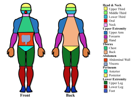
Select the region of the body you want to reconstruct with a local option or to serve as a donor site for reconstruction elsewhere in the body as a regional or free flap.
Head & Neck[edit | edit source]
Upper Third of Face[edit | edit source]
Middle Third of Face[edit | edit source]
Lower Third of Face[edit | edit source]
Oral[edit | edit source]
Neck[edit | edit source]
Upper Extremity[edit | edit source]
Upper Arm[edit | edit source]
Forearm[edit | edit source]
Hand[edit | edit source]
Thorax[edit | edit source]
Chest[edit | edit source]
Back[edit | edit source]
Abdomen[edit | edit source]
Abdominal Wall[edit | edit source]
Viscera[edit | edit source]
Perineum[edit | edit source]
Anterior[edit | edit source]
Posterior[edit | edit source]
Lower Extremity[edit | edit source]
Upper Leg[edit | edit source]
Lower Leg[edit | edit source]
Foot[edit | edit source]
Non-Regional[edit | edit source]
Random Pattern Flaps[edit | edit source]
Self assessment[edit | edit source]
- Review the contents of this page as well as the rest of the pages related to the Flaps & Classification section then go to the AmoSmile App to take the Self-Assessment quiz on Flaps & Classifications.
- See Z-Plasty Module navigation page for app download instructions.
