Fourier Transform Infrared Spectroscopy (FTIR) is an analytical technique used to identify chemical compounds and functional groups, by measuring how a sample absorbs infrared radiation at different wavelengths.
Molecules vibrate in characteristic ways when exposed to infrared light. Different chemical bonds absorb energy at specific frequencies, producing an infrared spectrum that acts like a molecular fingerprint.
Normalization adjusts spectra to a common scale so samples can be compared directly. Normalization helps to compensate for factors like different sample thickness, instrument variability, differences in crystal contact, and variations in sample concentration. After normalization, it will be much easier to compare peak heights and shapes between samples.
Raw FTIR spectra often have sloping or curved baselines due to instrument drift, scattering effects, or environmental interference. Baseline correction removes these artifacts so peak intensities more accurately reflect chemical composition.
There are many openly accessible softwares available for download on the internet which aid in the steps toward full analysis of a spectra, along with a human contribution.
We will explore 2 of these applications in detail on this page: Open Specy and Ira FTIR Explorer.
There are strengths and weaknesses to both which will be examined below, as well as steps to use.
Additionally, here are some other softwares which may be worth checking out but which are not thoroughly described on this page:
Quasar: An open-source spectroscopy analysis platform built on Python that integrates with the Orange data-mining environment. Quasar enables users to create reproducible workflows for spectral preprocessing, including baseline correction, normalization, smoothing, peak detection, and multivariate statistical analyses such as principal component analysis (PCA). It is particularly well suited for researchers processing large datasets or developing automated analysis pipelines.
SpectraFit: An open-source Python application designed specifically for interactive and automated spectral peak fitting. It provides tools for baseline correction, deconvolution of overlapping peaks, peak assignment, and quantitative curve fitting using common mathematical models (e.g., Gaussian, Lorentzian, and Voigt functions). SpectraFit is especially useful for obtaining accurate peak positions, widths, and intensities for detailed spectral analysis.
Spectragryph: A free spectroscopy analysis program for educational and non-commercial use that supports a wide range of spectroscopic techniques, including FTIR, Raman, UV-Vis, fluorescence, and NIR spectroscopy. It provides an intuitive graphical interface with tools for baseline correction, smoothing, normalization, peak picking, peak labeling, spectral subtraction, and overlaying multiple spectra for comparison. Spectragryph also includes spectral library searching, making it one of the most user-friendly options for identifying functional groups and comparing unknown samples with reference spectra.
Open Specy is a free, open-source software platform for analyzing Raman and FTIR spectra. Originally developed for identifying environmental plastics and microplastics, it has expanded into a general-purpose spectroscopy tool capable of preprocessing, visualizing, comparing, and identifying unknown spectra using an extensive community-maintained reference library. Open Specy is available as an online web application: https://www.openanalysis.org/openspecy/
How to Use
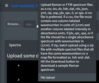
Export your FTIR data as a CSV, JDX, SPC, SPA, or ASP.
Navigate to the Open Specy website, and create an account if desired.
Select "Browse" and choose your exported file to upload it to the software. The spectrum should immediately appear on an interactive graph where users can zoom, pan, inspect peaks, and overlay multiple spectra.
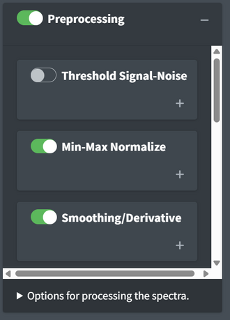
There are many options for preprocessing, the first three of which can be seen in the image. Options include normalization, smoothing, intensity adjustment, conform wavenumbers, baseline correction, range selection, flatten region, collapse particle spectra, and spatial smooth. Toggle these options on and off for different levels of processing and decide on the settings of choice before beginning any analysis.
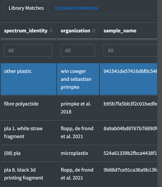
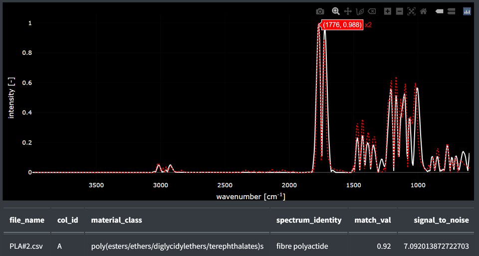
Turn on identification, and press more to see possible molecule matches to the spectra from the Open Specy library. Beside each match, the percent match is displayed. It is also possible to overlay the unknown and the reference spectrum.[[File:Identification.png|center|thumb|300x300px|[[File:Cross compare.png|center|thumb|]]]]
The software suggests likely compounds, but it is always a good idea for the user to identify major peak positions, peak intensities, functional groups, and whether the suggested match makes sense chemically. Spectral matching should be considered an aid rather than definitive proof of compound identity.
Users can export processed spectra after done with analysis.
Strengths
Free and open source
No programming required for the web version
Supports multiple FTIR and Raman file formats
Community-maintained spectral library
Automatic spectral matching
Weakesses
Reference library is strongest for polymers and environmental materials
Automatic matches should always be manually verified
Ira FTIR Explorer (Ira) is a free, open-source desktop application developed specifically for rapid visualization and analysis of FTIR spectra. Unlike Open Specy, which focuses heavily on automated library matching, Ira emphasizes interactive exploration of spectra. It provides tools for plotting, comparing, stacking, annotating, and detecting peaks within multiple spectra simultaneously.
Extract the downloaded files if necessary and launch the program
How to Use
Export your FTIR data as a CSV or TXT.
Select the file folder and choose the file you would like to upload to the software. The graph should be displayed immediately.
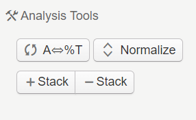
Use these buttons to switch from absorbance to % transmittance and to normalize. The stack + and - buttons can be used if you have uploaded more than one spectra. Pressing + will move the spectra closer together, while pressing - will separate them further.
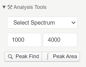
To detect peaks, select "Peak Detection" and the software will automatically identify the local maxima and label the peak position within a certain range of wavenumbers as specified by the user. It is possible to adjust detection parameters to avoid identifying noise as true peaks.
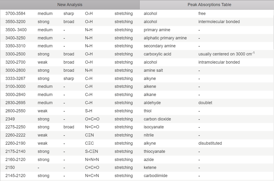
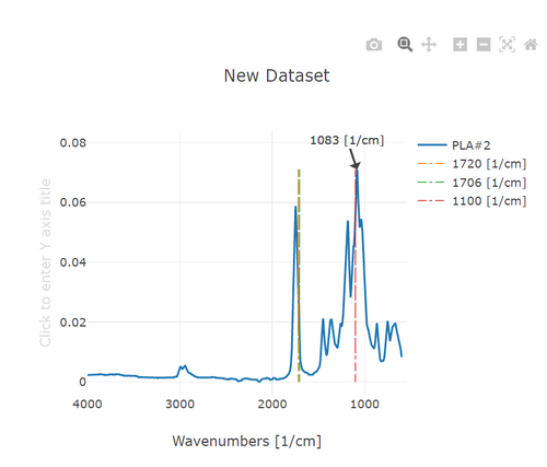
There is a Peak Absorption Table with a long list of common peak wavenumbers and what functional groups they represent. If the user selects one on the table, a line will appear at that wavenumber on the graph so it can be compared to peaks present in the spectra.
In addition to adding labels at pre-saved wavenumber values by pressing them on the table, users can manually add their own labels by inputting a wavenumber.