This page is part of the paramedic curriculum and is currently under development. Content will be added to this page, but there is no public knowledge currently available from this page. We thank you for you for your visit and hope you return when this program has been completed. Should you be interested in the foundation of all basic paramedic skills, click here to return to the EMT NREMT Skillset home page. For other sustainable projects, see Appropedia's home page.
This page is intended to provide working surface knowledge of the heart's anatomy and physiology. Keep in mind that both cardiac and systemic A&P are incredibly information dense topics and that there are doctoral fields of study for each. This page will give an overview of the cardiac system that is sufficient for the majority of paramedics, but it is always recommended to perform your own independent research and study as this page will not cover more in depth topics that may be salient to a paramedic's practice.
Anatomy of the Heart[edit | edit source]
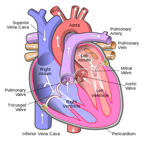
Walls of the Heart[edit | edit source]
Endocardium
Myocardium
Pericardium
Chambers of the Heart[edit | edit source]
Atria
Ventricles
Valves of the Heart[edit | edit source]
There are four valves of the heart listed in order as the tricuspid, pulmonary semilunar, mitral (bicuspid), and aortic semilunar valves. There is a mnemonic to easily remember these valves and their order: Toilet Paper My Assets. The valves are attached to chordae tendineae and papillary muscles that allow for closure during systole. Malformations or stenosis of valves may cause different heart tones and conditions such as regurgitation; these will not be discussed on this page as they are more in depth topics than a general overview of heart anatomy and physiology will cover.
Arteries and Veins of the Heart[edit | edit source]
Inferior and Superior Vena Cava
Pulmonary Arteries
Pulmonary Veins
Aorta
Coronary Arteries
- Left main coronary artery
- Left circumflex coronary artery
- Left anterior descending coronary artery
- Right coronary artery
- Posterior circulation and dominance
The Flow of Blood[edit | edit source]
This section will show the flow of blood through cardiac circulation starting at the superior and inferior vena cava. If you have a model or picture of the heart available, you may follow along for ease of visualization.
Blood initially enters cardiac circulation from the superior and inferior vena cava where it pours into the right atrium. As the atria contract, the blood is forced through the tricuspid valve into the right ventricle. The ventricles contract and the blood moves through the pulmonary semilunar valve into the pulmonary trunk of the pulmonary arteries. From there, blood goes through the lungs and is oxygenated in the pulmonary circulatory system before returning to the heart via the pulmonary veins. The pulmonary veins feed into the left atria, and when the atria contract the blood is now forced through the mitral (bicuspid) valve into the left ventricle. From the left ventricle, blood is moved through the aortic semilunar valve into the aorta where it either continues into systemic circulation or "falls back against" the aortic semilunar valve and flows into the coronary arteries to feed the cardiac muscle. The blood is transmitted by the arterial system to a location in the body and exchanges its oxygen for byproducts of cellular metabolism before moving back to the heart via the venous system (of which the last stop is either the superior or inferior vena cava).
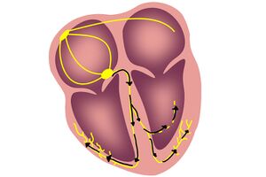
Electrical Conduction System[edit | edit source]
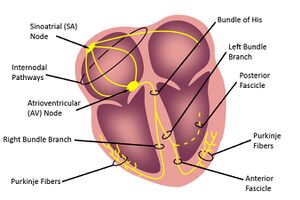
Sinoatrial Node[edit | edit source]
Internodal Pathways
Bachmann's Bundle
Atrioventricular Node/AV Junction[edit | edit source]
Bundle of His
Bundle Branches
Left Anterior and Posterior Fascicles
Purkinje Fibers[edit | edit source]
Cardiac Contraction[edit | edit source]
Physiology of the Heart[edit | edit source]
Important Atoms/Ions[1][edit | edit source]
Sodium (Na+)
Potassium (K+)
Chlorine (Cl-)
Calcium (Ca2+)
Magnesium (Mg2+)
Electrical Conduction[edit | edit source]
Muscular Contraction[edit | edit source]
The Heart in EMS[edit | edit source]
EKG Waveforms[edit | edit source]
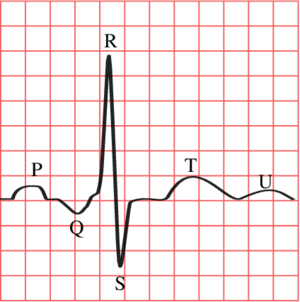
Isoelectric Line
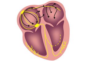
P wave
PR interval
QRS complex
J point
T wave
ST segment
U wave
QT interval
RR interval
The Bundle of Kent and WPW[edit | edit source]
- ↑ Grant, A. O. (2008). Cardiac Ion Channels. AHA Journals. Retrieved October 18, 2022, from https://www.ahajournals.org/doi/full/10.1161/CIRCEP.108.789081