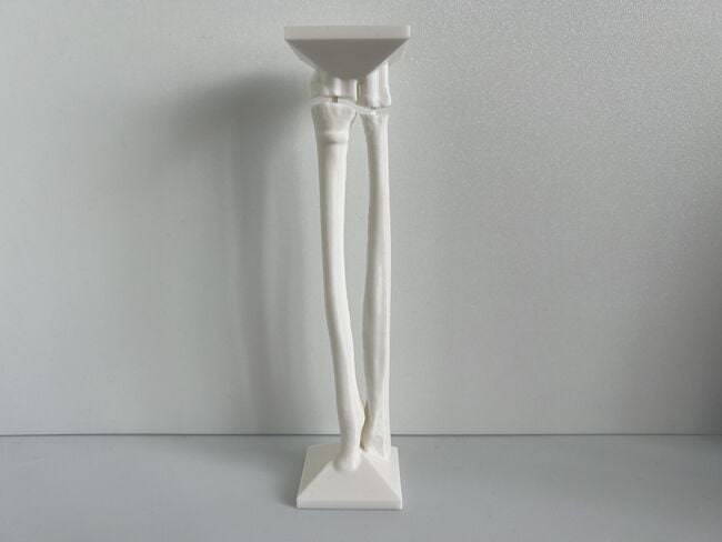
This module allows traditional bone setters, pre-hospital providers, clinical officers, nurses, nurse practitioners, and medical officers to become confident and competent in performing point-of-care ultrasound diagnostic imaging to rule out the presence of a pediatric distal forearm fracture and distinguish between buckle (torus) fractures and cortical break fractures to make appropriate referrals as part of the management of closed pediatric (< 16 years of age) distal forearm fractures in regions without access to X-ray imaging and orthopedic specialist coverage.[1][2][3][4][5][6][7][8][9]
Materials and Equipment[edit | edit source]
| # | Item | Image |
|---|---|---|
| 1 | 3D Printed Pediatric Forearm Simulator Mould | 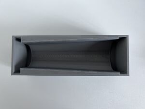 |
| 2 | Large Plate or Tray | 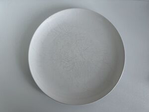 |
| 3 | Cellophane Wrap | 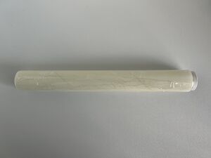 |
| 4 | 3D Printed Pediatric Female Forearm Bone Models (White PLA) #1-#6 for Unblinded Training | 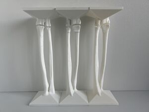 |
| 5 | 3D Printed Pediatric Female Forearm Bone Models (Black PLA) #5-#10 for Blinded Training | 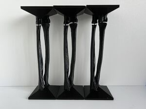 |
| 6 | Large Measuring Cup | |
| 7 | Water (~800 mL per mould pour) | |
| 8 | Supplies for Heating Water
|
|
| 9 | Weigh Scale | 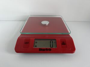 |
| 10 | Unflavoured (Clear) Gelatine (10 g per 100 mL of water, ~80 g per mould pour) | 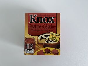 |
| 11 | Spoon | |
| 12 | Blue, Red and Yellow Food Colouring or Black Food Colouring | 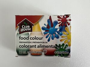 |
| 13 | Refrigerator | |
| 14 | Alarm Timer | |
| 15 | Knife | |
| 16 | Needle Nose Pliers | |
| 17 | Toothbrush | |
| 18 | Mild Detergent | |
| 19 | Sink |
Preparing Pediatric Forearm Simulators for Unblinded Training[edit | edit source]
- Place mould on a large plate or tray in case of liquid spillage.
- Line the interior of the mould with clear cellophane wrap.
- Place a white PLA 3D Printed Pediatric Female Forearm Bone Model into the mould. The arrows of the base of the bone models should be aligned with the arrows on the ends of the mould. The letter "R" (which stands for radius) on the base of each bone model should align with the letter "R" on the end of the mould and the letter "U" (which stands for ulna) on the base of each bone model should align with the letter "U" on the end of the mould.
- Heat up 400 mL of water in a kettle, on a stove, or in a microwave.
- Add 80 grams of unflavored gelatine to a large measuring cup filled with 400 mL of water at room temperature.
- Mix well to remove any solid masses.
- Add the heated 400 mL of water to the gelatine solution.
- Mix well so all the gelatine dissolves.
- Pour the gelatine solution into the mould.
- Carefully place the mould in the refrigerator while avoiding spillage.
- Wait 30 minutes for the gelatine to settle into the mould and then pour the leftover gelatine solution to fill up the mould.
- Refrigerate the mould for 4 hours to completely set.
- Gently slide a knife in between the mould and cellophane to help loosen the mould.
- While an assistant stabilizes the mould with both hands on a flat surface, grab the base of each bone model with needle nose pliers and carefully extract the Pediatric Forearm Simulator from the mould.
- Remove the cellophane from the Pediatric Forearm Simulator.
- After use, gently wipe off the ultrasound gel on the Pediatric Forearm Simulator and store the Pediatric Forearm Simulator in a refrigerator.
- Use cold water, mild detergent and toothbrush to clean the 3D Printed Pediatric Forearm Simulator Mould and 3D Printed Pediatric Female Forearm Bone Model for reuse.
Preparing Pediatric Forearm Simulators for Blinded Training[edit | edit source]
- Place mould on a large plate or tray in case of liquid spillage.
- Line the interior of the mould with clear cellophane wrap.
- Place a black PLA 3D Printed Pediatric Female Forearm Bone Model into the mould. The arrows of the base of the bone models should be aligned with the arrows on the ends of the mould. The letter "R" (which stands for radius) on the base of each bone model should align with the letter "R" on the end of the mould and the letter "U" (which stands for ulna) on the base of each bone model should align with the letter "U" on the end of the mould.
- Heat up 400 mL of water in a kettle, on a stove, or in a microwave.
- Add 80 grams of unflavored gelatine to a large measuring cup filled with 400 mL of water at room temperature.
- Mix well to remove any solid masses.
- Add the heated 400 mL of water to the gelatine solution.
- Mix well so all the gelatine dissolves.
- **Add 40 drops of blue food colouring, 20 drops red food colouring, and 20 drops yellow food colouring or 80 drops of black food colouring and mix well.**
- Pour the gelatine solution into the mould.
- Carefully place the mould in the refrigerator while avoiding spillage.
- Wait 30 minutes for the gelatine to settle into the mould and then pour the leftover gelatine solution to fill up the mould.
- Refrigerate the mould for 4 hours to completely set.
- Gently slide a knife in between the mould and cellophane to help loosen the mould.
- While an assistant stabilizes the mould with both hands on a flat surface, grab the base of the bone model with needle nose pliers and carefully extract the Pediatric Forearm Simulator from the mould.
- Remove the cellophane from the Pediatric Forearm Simulator.
- After use, gently wipe off the ultrasound gel on the Pediatric Forearm Simulator and store the Pediatric Forearm Simulator in a refrigerator.
- Use cold water, mild detergent and toothbrush to clean the 3D Printed Pediatric Forearm Simulator Mould and 3D Printed Pediatric Female Forearm Bone Model for reuse.
Do not use a dishwasher to clean a 3D Printed Pediatric Forearm Simulator Mould or 3D Printed Pediatric Female Forearm Bone Model because the thermoplastic may deform when exposed to heated water.
Acknowledgements[edit | edit source]
This work is funded by a grant from the Intuitive Foundation. Any research, findings, conclusions, or recommendations expressed in this work are those of the author(s), and not of the Intuitive Foundation.
References[edit | edit source]
- ↑ Onyemaechi NO, Itanyi IU, Ossai PO, Ezeanolue EE. Can traditional bonesetters become trained technicians? Feasibility study among a cohort of Nigerian traditional bonesetters. Hum Resour Health. 2020 Mar 20;18(1):24. doi: 10.1186/s12960-020-00468-w. PMID: 32197617; PMCID: PMC7085192.
- ↑ Heiner JD, McArthur TJ. The ultrasound identification of simulated long bone fractures by prehospital providers. Wilderness Environ Med. 2010 Jun;21(2):137-40. doi: 10.1016/j.wem.2009.12.028. Epub 2009 Dec 22. PMID: 20591377.
- ↑ Heiner JD, Baker BL, McArthur TJ. The ultrasound detection of simulated long bone fractures by U.S. Army Special Forces Medics. J Spec Oper Med. 2010 Spring;10(2):7-10. PMID: 20936597.
- ↑ Heiner JD, Proffitt AM, McArthur TJ. The ability of emergency nurses to detect simulated long bone fractures with portable ultrasound. Int Emerg Nurs. 2011 Jul;19(3):120-4. doi: 10.1016/j.ienj.2010.08.004. Epub 2010 Sep 25. PMID: 21665155.
- ↑ Snelling PJ, Jones P, Keijzers G, Bade D, Herd DW, Ware RS. Nurse practitioner administered point-of-care ultrasound compared with X-ray for children with clinically non-angulated distal forearm fractures in the ED: a diagnostic study. Emerg Med J. 2021 Feb;38(2):139-145. doi: 10.1136/emermed-2020-209689. Epub 2020 Sep 8. PMID: 32900856.
- ↑ Snelling PJ, Jones P, Moore M, Gimpel P, Rogers R, Liew K, Ware RS, Keijzers G. Describing the learning curve of novices for the diagnosis of paediatric distal forearm fractures using point-of-care ultrasound. Australas J Ultrasound Med. 2022 Mar 7;25(2):66-73. doi: 10.1002/ajum.12291. PMID: 35722050; PMCID: PMC9201201.
- ↑ Heiner JD, McArthur TJ. A simulation model for the ultrasound diagnosis of long-bone fractures. Simul Healthc. 2009 Winter;4(4):228-31. doi: 10.1097/SIH.0b013e3181b1a8d0. PMID: 19915442.
- ↑ Snelling PJ, Keijzers G, Byrnes J, Bade D, George S, Moore M, Jones P, Davison M, Roan R, Ware RS. Bedside Ultrasound Conducted in Kids with distal upper Limb fractures in the Emergency Department (BUCKLED): a protocol for an open-label non-inferiority diagnostic randomised controlled trial. Trials. 2021 Apr 14;22(1):282. doi: 10.1186/s13063-021-05239-z. PMID: 33853650; PMCID: PMC8048294.
- ↑ Snelling PJ. A low-cost ultrasound model for simulation of paediatric distal forearm fractures. Australas J Ultrasound Med. 2018 Feb 25;21(2):70-74. doi: 10.1002/ajum.12083. PMID: 34760505; PMCID: PMC8409885.
