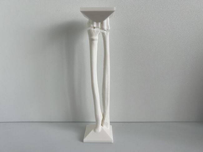
This module allows traditional bone setters, pre-hospital providers, clinical officers, nurses, nurse practitioners, and medical officers to become confident and competent in performing point-of-care ultrasound diagnostic imaging to rule out the presence of a pediatric distal forearm fracture and distinguish between buckle (torus) fractures and cortical break fractures to make appropriate referrals as part of the management of closed pediatric (< 16 years of age) distal forearm fractures in regions without access to X-ray imaging and orthopedic specialist coverage.[1][2][3][4][5][6][7][8][9] These 3D printed bone models feature a semi-engraved model number, gender symbol, and two mould insertion direction arrows on the base of each model to assist with model identification and proper orientation of each bone model in the mould.
Lessons Learned[edit | edit source]
We provide information on how lessons learnt from the development of our original prototype surgical module have been used in the development of this additional sub-module.
Development of Simulator[edit | edit source]
We provide information on how lessons learnt from the development of our original prototype surgical module have been used in the development of the simulator for this additional sub-module.
3D Printed Pediatric Female Forearm Bone Models[edit | edit source]
In partnership with Qrint Studio, our team rapidly prototyped the 3D printed pediatric female forearm bone models after the August 25-26, 2022 site visit using the design choices outlined below:
- We re-scaled the forearm bone models so the total length of the radius is 182.84 mm (185.39 mm - 2.55 mm) so the exported stl file can be printed at 100% scale and the simulator will be 185.39 mm long when the male connector parts with a 2.55 mm length offset are inserted into the female receptive parts.[10][11]
- We adjusted the position of the female part in the bone models so the opening is in a consistent position for all the bone models and is not too close to the edge which would result in a hole in the bone's 3D printed outer surface.
- We printed multiple iterations, observed shrinkage of the female parts, and tested the male connector parts to identify the optimal printer resolution (0.20 mm), male part tolerance (0.25 mm) and suitable dimensions. We increased the depth of the female parts so the male parts can be inserted into the female parts with a minimal amount of force and will stay in place once inserted.
- We rounded off the distal epiphysis and proximal metaphysis more so the edges are curved and smoothed out the metaphysis of the radius and ulna for a more realistic and improved aesthetic appearance of the bone models.
- We revised the square base so each identical side of the forearm bone base is at least 7 cm long instead of 5 cm. A longer base projects more out of the top of the open face mould to permit easier removal of the Pediatric Forearm Simulator after the gelatine pour.
- We positioned the forearm bone model in the central part of the base so the forearm soft tissue is not too close to the edge of the base which would result in inadequate gelatine coverage and potential exposure of the 3D printed bone model. A simulated soft tissue layer overlying the 3D printed bone simulation model is required for ultrasound training.
- We added semi-engraved numbers in Arabic, English, and Chinese characters for the 6 official languages of the United Nations, female gender icon, our team logo and mould placement direction arrows in the base of each model. Each proximal forearm bone model is labelled with an odd number and each distal forearm bone model is labelled with an even number. We printed three model base iterations and identified an ideal semi-engraving depth of 4 mm which optimizes shadow contrast and visibility of the semi-engraved features.
- To avoid print failures, we recommend printing each Pediatric Female Forearm Bone Model component separately and not simultaneously on the same 3D printer.
- Based on our prototyping, we recommend using only one connector in the ulna for the No Fracture Model to permit the learner to visualize the physis without acoustic interference from the radiopaque connector. However, the connectors for the other models are thinner because the female parts have to be a smaller diameter in order to avoid printing issues on the outer surface of the bone model. Therefore, we recommend using both connectors for the radius and ulna to optimize the stability of the model, especially during the removal of the simulator from the mould. We recommend including extra sets of connectors in case they are broken and need to be replaced.
- We noticed that the prints had a suboptimal appearance when sliced with Ultimaker Cura for a non-Ultimaker printer. This may be due to the fact that Ultimaker Cura Generic PLA settings are optimized for Ultimaker and not Prusa i3MK3S printers. Initially, we slowed down the print speed to 40 mm/s and top/bottom layer speed to 5 mm/s to improve the quality of the printed models. However, this substantially increased the print time.
- We tested the default settings of the slicing program recommended by the printer manufacturer (i.e., using Prusa Slicer for Prusa i3MK3S printer instead of Ultimaker Cura slicer program) and found that this required minimal inputs to the slicing program, gave prints of acceptable quality, and kept the print times under 8 hours.
- We also identified an issue with the base of Model 6 not being flush with the build plate which was leading to print failures. We imported the stl files into Simplify3D to correct this issue.
- We recommend using a spatula to remove the bone models from a non-flexible build plate to prevent breakage.
3D Printed Pediatric Female Forearm Simulator Mould[edit | edit source]
In partnership with Qrint Studio, our team rapidly prototyped the reusable, 3D printed pediatric female forearm simulator moulds after the August 25-26, 2022 site visit using the design choices outlined below:
- We re-designed the 3D Printed Pediatric Female Forearm Simulator Mould with concave cavity dimensions based on previously published forearm and wrist circumference values for the left upper extremity of 10 year old females.[12][13]
- Although the distal forearm bones are very superficial, we repositioned the forearm bones in the mould in order to have adequate simulated soft tissue coverage for simulation training.
- We increased the tolerance of the concave cavity of the negative mould to permit easier extraction of the Pediatric Forearm Simulator from the mould after the gelatin pour. The added benefit of this tolerance is it permits the forearm bones to fit inside the mould, even if the mould is warped slightly during the 3D printing process since PLA filament is prone to warping.
- We added a semi-engraved female gender icon, our team logo, "R" and "U" initials (corresponding to the radius and ulna) and bone model insertion direction arrows at the end of the mould to assist with proper orientation of each bone model in the mould.
- The reusable mould is compatible with all 3D Printed Pediatric Female Forearm Bone Models #1-#10 so only 1-2 moulds are required which significantly reduces simulator costs and production time.
- Based on user feedback, we wanted to minimize any required custom print settings for our mould. This mould can be printed using default settings for PLA filament except for build plate temperature which should be confirmed to be set to a minimum of 55 degrees Celsius. Changing the build plate temperature can be performed using any 3D printer's slicing software and does not require using Ultimaker Cura or Cura Lulzbot slicing software.
- Based on user feedback from 3D printing organizations, we included ready-to-print g-codes that can be downloaded from a Google drive and Appropedia and stl files to permit slicing by individuals with access to 3D printers. One 3D printing organization informed us that Appropedia's workaround on how to save g-code files uploaded to Appropedia was too foreign and these g-codes would be not be downloaded and saved despite the availability of written instructions. Thus, we made sure that the g-code files would be downloaded from a Google drive and that stl files could be downloaded from Appropedia to permit slicing for the locally available 3D printer.
Development of Educational Material[edit | edit source]
We provide information on how lessons learnt from the development of our original prototype surgical module have been used in the development of the educational material for this additional sub-module.
On September 3, 2022, we spoke with 5 traditional bone setters about our prototype open-access, and self-assessed simulation training module on ultrasound diagnosis of pediatric distal forearm fractures.
We created over 25 innovative digital illustrations to empower them to learn the anatomy terms required for ultrasound diagnosis of pediatric distal forearm fractures.
The positive feedback we received on our aviation-style procedural checklists for our original prototype surgical module led us to apply a similar methodology for this educational material for the skills training module.
Development of Self-Assessment Framework[edit | edit source]
We provide information on how lessons learnt from the development of our original prototype surgical module have been used in the self-assessment portion of this additional sub-module.
The positive feedback we received on our aviation-style procedural checklists for our original prototype surgical module led us to apply a similar methodology for this self-assessment framework for this module.
Our self-assessment framework includes self-review of labelled images because the mobile apps for portable ultrasound devices permit the labelling and emailing of images for self-assessment. The positive feedback we received on our quality assurance checklist of our 3D printed bone models led us to apply a similar strategy for learner self-review of their labelled ultrasound images. This methodology enables the learner to:
- Ensure they are practicing the appropriate skills
- Modify their performance to improve competence; and
- Determine when they have practiced to a sufficient level of mastery to perform the procedure in a patient.
Acknowledgements[edit | edit source]
This work is funded by a grant from the Intuitive Foundation. Any research, findings, conclusions, or recommendations expressed in this work are those of the author(s), and not of the Intuitive Foundation.
References[edit | edit source]
- ↑ Onyemaechi NO, Itanyi IU, Ossai PO, Ezeanolue EE. Can traditional bonesetters become trained technicians? Feasibility study among a cohort of Nigerian traditional bonesetters. Hum Resour Health. 2020 Mar 20;18(1):24. doi: 10.1186/s12960-020-00468-w. PMID: 32197617; PMCID: PMC7085192.
- ↑ Heiner JD, McArthur TJ. The ultrasound identification of simulated long bone fractures by prehospital providers. Wilderness Environ Med. 2010 Jun;21(2):137-40. doi: 10.1016/j.wem.2009.12.028. Epub 2009 Dec 22. PMID: 20591377.
- ↑ Heiner JD, Baker BL, McArthur TJ. The ultrasound detection of simulated long bone fractures by U.S. Army Special Forces Medics. J Spec Oper Med. 2010 Spring;10(2):7-10. PMID: 20936597.
- ↑ Heiner JD, Proffitt AM, McArthur TJ. The ability of emergency nurses to detect simulated long bone fractures with portable ultrasound. Int Emerg Nurs. 2011 Jul;19(3):120-4. doi: 10.1016/j.ienj.2010.08.004. Epub 2010 Sep 25. PMID: 21665155.
- ↑ Snelling PJ, Jones P, Keijzers G, Bade D, Herd DW, Ware RS. Nurse practitioner administered point-of-care ultrasound compared with X-ray for children with clinically non-angulated distal forearm fractures in the ED: a diagnostic study. Emerg Med J. 2021 Feb;38(2):139-145. doi: 10.1136/emermed-2020-209689. Epub 2020 Sep 8. PMID: 32900856.
- ↑ Snelling PJ, Jones P, Moore M, Gimpel P, Rogers R, Liew K, Ware RS, Keijzers G. Describing the learning curve of novices for the diagnosis of paediatric distal forearm fractures using point-of-care ultrasound. Australas J Ultrasound Med. 2022 Mar 7;25(2):66-73. doi: 10.1002/ajum.12291. PMID: 35722050; PMCID: PMC9201201.
- ↑ Heiner JD, McArthur TJ. A simulation model for the ultrasound diagnosis of long-bone fractures. Simul Healthc. 2009 Winter;4(4):228-31. doi: 10.1097/SIH.0b013e3181b1a8d0. PMID: 19915442.
- ↑ Snelling PJ, Keijzers G, Byrnes J, Bade D, George S, Moore M, Jones P, Davison M, Roan R, Ware RS. Bedside Ultrasound Conducted in Kids with distal upper Limb fractures in the Emergency Department (BUCKLED): a protocol for an open-label non-inferiority diagnostic randomised controlled trial. Trials. 2021 Apr 14;22(1):282. doi: 10.1186/s13063-021-05239-z. PMID: 33853650; PMCID: PMC8048294.
- ↑ Snelling PJ. A low-cost ultrasound model for simulation of paediatric distal forearm fractures. Australas J Ultrasound Med. 2018 Feb 25;21(2):70-74. doi: 10.1002/ajum.12083. PMID: 34760505; PMCID: PMC8409885.
- ↑ Ng L, Saul T, Lewiss RE. Sonographic baseline physeal plate width measurements in healthy, uninjured children. Pediatr Emerg Care. 2014 Dec;30(12):871-4. doi: 10.1097/PEC.0000000000000290. PMID: 25407037.
- ↑ Gindhart PS. Growth standards for the tibia and radius in children aged one month through eighteen years. Am J Phys Anthop 1973; 39(1): 41–8.
- ↑ Edmond T, Laps A, Case AL, O'Hara N, Abzug JM. Normal Ranges of Upper Extremity Length, Circumference, and Rate of Growth in the Pediatric Population. Hand (N Y). 2020 Sep;15(5):713-721. doi: 10.1177/1558944718824706. Epub 2019 Feb 1. PMID: 30709325; PMCID: PMC7543216.
- ↑ Öztürk A, Çiçek B, Mazıcıoğlu MM, Zararsız G, Kurtoğlu S. Wrist Circumference and Frame Size Percentiles in 6-17-Year-Old Turkish Children and Adolescents in Kayseri. J Clin Res Pediatr Endocrinol. 2017 Dec 15;9(4):329-336. doi: 10.4274/jcrpe.4265. Epub 2017 May 17. PMID: 28515034; PMCID: PMC5785639.
