
Pediatric Distal Forearm Fractures/Ultrasound Diagnosis of Pediatric Distal Forearm Fractures
| Part of | Pediatric Distal Forearm Fractures |
|---|
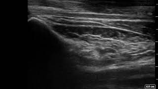
_Fracture_of_Pediatric_Female_Left_Distal_Radius_v2.0.jpg)
This module allows traditional bone setters, pre-hospital providers, clinical officers, nurses, nurse practitioners, and medical officers to become confident and competent in performing point-of-care ultrasound diagnostic imaging to rule out the presence of a pediatric distal forearm fracture and distinguish between buckle (torus) fractures and cortical break fractures to make appropriate referrals as part of the management of closed pediatric (< 16 years of age) distal forearm fractures in regions without access to X-ray imaging and orthopedic specialist coverage.[1][2][3][4][5][6][7][8][9]
Skills Training Objectives
[edit | edit source]- Identify the primary sonographic features of no fracture, a buckle (torus) fractures and a cortical break fracture of the pediatric distal forearm
- Describe the secondary sonographic features of a cortical break fracture of the pediatric distal forearm
- Perform the 6 view method for ultrasonographic diagnosis of pediatric distal forearm fractures
- Practise conducting a bilateral ultrasound scan to diagnose a pronator quadratus hematoma
Materials and Equipment
[edit | edit source]| # | Item |
|---|---|
| 1 | Any ultrasound device with a linear transducer (from 3.5 to 16 MHz and more) is suitable for fracture sonography. Nearly almost all commercially available portable ultrasound devices can be used. |
| 2 | Mobile device with ultrasound app installed |
| 3 | Ultrasound gel |
| 4 | 3 Pediatric Forearm Simulators for unblinded training (White 3D Printed Pediatric Female Bone Models #1-6) |
| 5 | 3 Pediatric Forearm Simulators for blinded training (Black 3D Printed Pediatric Female Bone Models #5-10) |
| 6 | Training Logbook |
| 7 | Self-Assessment Framework |
| 7 | Clipboard and pen |
Basic Ultrasound Skills Training
[edit | edit source]Practice basic ultrasound skills on each other's arms:
- Fanning the iQ
- Sliding the iQ
- Rotating the iQ
- Proper Hand Positioning
- Choosing the Right Preset
- Adjusting the Overall Gain
6 View Technique for Ultrasound Scanning of Pediatric Distal Forearm Fractures
[edit | edit source]Practice the 6 view technique for ultrasound scanning of pediatric distal forearm fractures on each other's arms. Position the forearm so it is resting comfortably on a pillow, towel or examination bed.
Apply copious amount of ultrasound gel to the distal forearm.
Start by scanning the dorsal aspect of the radius without applying pressure on the injured area and by gently resting the probe on the gel.

Align probe (black) with the longitudinal axis of the bone with the probe marker (blue) oriented distally. Start scanning at the mid-shaft and carefully move down to the epiphysis.

Acquire, label, and save an image of the epiphysis (E), metaphysis (M), and diaphysis (D).

Repeat steps 2,4 and 5 for the dorsal aspect of the ulna.

Repeat steps 2,4 and 5 for the lateral aspect of the ulna.

Repeat steps 2,4 and 5 for the lateral aspect of the radius.

Gently supinate the forearm to at least 90 degrees to access the volar (palmar) aspect of the distal forearm.

Repeat steps 2,4 and 5 for the volar (palmar) aspect of the radius.

Repeat steps 2,4 and 5 for the volar (palmar) aspect of the ulna.
Repeat steps 2-11 on opposite forearm to identify asymmetric bends or breaks in the bone cortex.
Review all the acquired images to determine if they meet all the standards outlined in the quality assurance checklist in the self-assessment framework.
Ultrasound Scanning for Pronator Quadratus Hematoma Sign
[edit | edit source]Practise scanning the pronator quadratus muscle on each other's arms.

With the forearm supinated at last 90 degrees, align the probe with the metaphysis of the distal radius and increase the depth to 4 cm.
Record a 4 second video slowly sweeping across the volar (palmar) aspect of the distal forearm while maintaining the probe perpendicular to the skin. Sweep until the metaphysis of the distal ulna is in the field of view.

Review the video to obtain an image of the largest section of the pronator quadratus muscle, which is typically the mid-portion of the sweep.

Repeat steps 1-3 with the opposite forearm for comparison. Compare the thickness and appearance of the pronator quadratus muscle on both sides.
Review all the acquired images to determine if they meet all the standards outlined in the quality assurance checklist in the self-assessment framework.
Unblinded Training on Pediatric Female Forearm Simulators
[edit | edit source]- Acquire, label, and save an image of the epiphysis, physis, metaphysis and diaphysis of the radius and ulna in each view.
- Identify a buckle fracture which appears as an asymmetric outward or inward bending of the bone cortex in each view.
- Identify a cortical break fracture which appears as a black zone in the bright, sharp white line of the bone cortex in each view.
- Determine if no fracture, a buckle fracture, or a cortical break fracture is present in each view.
- Select the appropriate treatment plan for each simulated patient.
- Fill out the training logbook.
- Review all the acquired images to determine if they meet all the standards outlined in the quality assurance checklist in the self-assessment framework.
Blinded Training on Pediatric Female Forearm Simulators
[edit | edit source]
- Acquire, label, and save an image of the epiphysis, physis, metaphysis and diaphysis of the radius and ulna in each view.
- Identify a buckle fracture which appears as an asymmetric outward or inward bending of the bone cortex in each view.
- Identify a cortical break fracture which appears as a black zone in the bright, sharp white line of the bone cortex in each view.
- Determine if no fracture, a buckle fracture, or a cortical break fracture is present in each view.
- Select the appropriate treatment plan for each simulated patient.
- Fill out and sign the training logbook.
- Review all the acquired images to determine if they meet all the standards outlined in the quality assurance checklist in the self-assessment framework.
Acknowledgements
[edit | edit source]This work is funded by a grant from the Intuitive Foundation. Any research, findings, conclusions, or recommendations expressed in this work are those of the author(s), and not of the Intuitive Foundation.
References
[edit | edit source]- ↑ Onyemaechi NO, Itanyi IU, Ossai PO, Ezeanolue EE. Can traditional bonesetters become trained technicians? Feasibility study among a cohort of Nigerian traditional bonesetters. Hum Resour Health. 2020 Mar 20;18(1):24. doi: 10.1186/s12960-020-00468-w. PMID: 32197617; PMCID: PMC7085192.
- ↑ Heiner JD, McArthur TJ. The ultrasound identification of simulated long bone fractures by prehospital providers. Wilderness Environ Med. 2010 Jun;21(2):137-40. doi: 10.1016/j.wem.2009.12.028. Epub 2009 Dec 22. PMID: 20591377.
- ↑ Heiner JD, Baker BL, McArthur TJ. The ultrasound detection of simulated long bone fractures by U.S. Army Special Forces Medics. J Spec Oper Med. 2010 Spring;10(2):7-10. PMID: 20936597.
- ↑ Heiner JD, Proffitt AM, McArthur TJ. The ability of emergency nurses to detect simulated long bone fractures with portable ultrasound. Int Emerg Nurs. 2011 Jul;19(3):120-4. doi: 10.1016/j.ienj.2010.08.004. Epub 2010 Sep 25. PMID: 21665155.
- ↑ Snelling PJ, Jones P, Keijzers G, Bade D, Herd DW, Ware RS. Nurse practitioner administered point-of-care ultrasound compared with X-ray for children with clinically non-angulated distal forearm fractures in the ED: a diagnostic study. Emerg Med J. 2021 Feb;38(2):139-145. doi: 10.1136/emermed-2020-209689. Epub 2020 Sep 8. PMID: 32900856.
- ↑ Snelling PJ, Jones P, Moore M, Gimpel P, Rogers R, Liew K, Ware RS, Keijzers G. Describing the learning curve of novices for the diagnosis of paediatric distal forearm fractures using point-of-care ultrasound. Australas J Ultrasound Med. 2022 Mar 7;25(2):66-73. doi: 10.1002/ajum.12291. PMID: 35722050; PMCID: PMC9201201.
- ↑ Heiner JD, McArthur TJ. A simulation model for the ultrasound diagnosis of long-bone fractures. Simul Healthc. 2009 Winter;4(4):228-31. doi: 10.1097/SIH.0b013e3181b1a8d0. PMID: 19915442.
- ↑ Snelling PJ, Keijzers G, Byrnes J, Bade D, George S, Moore M, Jones P, Davison M, Roan R, Ware RS. Bedside Ultrasound Conducted in Kids with distal upper Limb fractures in the Emergency Department (BUCKLED): a protocol for an open-label non-inferiority diagnostic randomised controlled trial. Trials. 2021 Apr 14;22(1):282. doi: 10.1186/s13063-021-05239-z. PMID: 33853650; PMCID: PMC8048294.
- ↑ Snelling PJ. A low-cost ultrasound model for simulation of paediatric distal forearm fractures. Australas J Ultrasound Med. 2018 Feb 25;21(2):70-74. doi: 10.1002/ajum.12083. PMID: 34760505; PMCID: PMC8409885.
| Authors | Julielynn Wong, Habila Umaru |
|---|---|
| License | CC-BY-SA-4.0 |
| Organizations | Medical Makers |
| Cite as | Medical Makers (2022–2026). "Pediatric Distal Forearm Fractures/Ultrasound Diagnosis of Pediatric Distal Forearm Fractures". Appropedia. Retrieved July 14, 2026. |