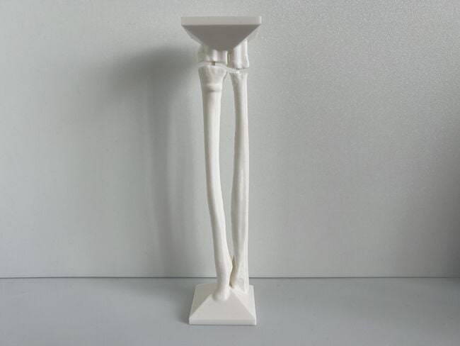
This module allows traditional bone setters, pre-hospital providers, clinical officers, nurses, nurse practitioners, and medical officers to become confident and competent in performing point-of-care ultrasound diagnostic imaging to rule out the presence of a pediatric distal forearm fracture and distinguish between buckle (torus) fractures and cortical break fractures to make appropriate referrals as part of the management of closed pediatric (< 16 years of age) distal forearm fractures in regions without access to X-ray imaging and orthopedic specialist coverage.[1][2][3][4][5][6][7][8][9]
Clinical Evaluation[edit | edit source]
Learning Objectives[edit | edit source]
- Conduct a comprehensive history and physical examination of a pediatric patient with a suspected distal forearm fracture
- Know the clinical indications and contraindications for using ultrasound as the sole imaging modality in the diagnosis of pediatric distal forearm fractures
- Know the indications for referral for pediatric distal forearm fractures
History[edit | edit source]
- Age
- Children < 12 years of age are more likely to sustain buckle fractures of the distal radius metaphysis
- Children > 12 years of age are more likely to sustain physeal fractures of the distal radius because the bone is stronger in comparison to the cartilaginous physis[10]
- Hand preference
- Injured arm
- Mechanism of injury
- Most are due to a fall on an outstretched hand (FOOSH). The scaphoid bone in these patients should be carefully evaluated.
- A direct strike to the forearm will inform the region of interest for scanning.
- A twisting mechanism is more likely to result in a soft tissue injury.
- Time of injury
- Previous forearm injury or birth defect
- Analgesia received
Physical Examination[edit | edit source]
Look[edit | edit source]
- The hand, forearm and elbow of the injured arm should be inspected for signs of swelling, bruising or deformity.
- Patients with visible deformity must be referred for x-ray imaging.
Feel[edit | edit source]
- The bones of the injured arm should be systematically palpated for tenderness or crepitus, being careful to gently examine any region of interest last.
- The scaphoid bone should be evaluated in children > 10 years of age by palpating the anatomical snuffbox and tubercle.
Move[edit | edit source]
- The full range of movement of the wrist should be evaluated as tolerated by the patient, which includes: flexion, extension, lateral movements (radius and ulna deviation), supination, pronation. Any pain or restriction of movement should be noted.
- Any significant pain or restriction of movement of the hand or elbow should prompt the patient being referred for x-ray imaging.
Neurovascular Exam[edit | edit source]
Clinical Indications for Ultrasound Scanning of a Pediatric Distal Forearm Injury[edit | edit source]
- Isolated, distal forearm injury with no visible deformity in a patient aged younger than 16 years[12]
Contraindications for Ultrasound Scanning of a Pediatric Distal Forearm Injury[edit | edit source]
If one or more of these symptoms are present, the pediatric patient with a distal forearm injury must be referred for X-ray imaging:
- Patient > 16 years of age
- Obvious angulation/deformity (soft tissue swelling allowed)
- Injury > 48 h old
- Bone disease (e.g. osteogenesis imperfecta)
- Congenital forearm malformation (e.g. radius hypoplasia)
- Compound/open fracture
- Neurovascular compromise
- Suspicion for other fracture (i.e., scaphoid or elbow fracture)
- Inability to clinically assess child
- Clinical suspicion e.g. ‘pain out of proportion’ despite normal ultrasound findings[12][13]
Additional Resources[edit | edit source]
Acknowledgements[edit | edit source]
This work is funded by a grant from the Intuitive Foundation. Any research, findings, conclusions, or recommendations expressed in this work are those of the author(s), and not of the Intuitive Foundation.
References[edit | edit source]
- ↑ Onyemaechi NO, Itanyi IU, Ossai PO, Ezeanolue EE. Can traditional bonesetters become trained technicians? Feasibility study among a cohort of Nigerian traditional bonesetters. Hum Resour Health. 2020 Mar 20;18(1):24. doi: 10.1186/s12960-020-00468-w. PMID: 32197617; PMCID: PMC7085192.
- ↑ Heiner JD, McArthur TJ. The ultrasound identification of simulated long bone fractures by prehospital providers. Wilderness Environ Med. 2010 Jun;21(2):137-40. doi: 10.1016/j.wem.2009.12.028. Epub 2009 Dec 22. PMID: 20591377.
- ↑ Heiner JD, Baker BL, McArthur TJ. The ultrasound detection of simulated long bone fractures by U.S. Army Special Forces Medics. J Spec Oper Med. 2010 Spring;10(2):7-10. PMID: 20936597.
- ↑ Heiner JD, Proffitt AM, McArthur TJ. The ability of emergency nurses to detect simulated long bone fractures with portable ultrasound. Int Emerg Nurs. 2011 Jul;19(3):120-4. doi: 10.1016/j.ienj.2010.08.004. Epub 2010 Sep 25. PMID: 21665155.
- ↑ Snelling PJ, Jones P, Keijzers G, Bade D, Herd DW, Ware RS. Nurse practitioner administered point-of-care ultrasound compared with X-ray for children with clinically non-angulated distal forearm fractures in the ED: a diagnostic study. Emerg Med J. 2021 Feb;38(2):139-145. doi: 10.1136/emermed-2020-209689. Epub 2020 Sep 8. PMID: 32900856.
- ↑ Snelling PJ, Jones P, Moore M, Gimpel P, Rogers R, Liew K, Ware RS, Keijzers G. Describing the learning curve of novices for the diagnosis of paediatric distal forearm fractures using point-of-care ultrasound. Australas J Ultrasound Med. 2022 Mar 7;25(2):66-73. doi: 10.1002/ajum.12291. PMID: 35722050; PMCID: PMC9201201.
- ↑ Heiner JD, McArthur TJ. A simulation model for the ultrasound diagnosis of long-bone fractures. Simul Healthc. 2009 Winter;4(4):228-31. doi: 10.1097/SIH.0b013e3181b1a8d0. PMID: 19915442.
- ↑ Snelling PJ, Keijzers G, Byrnes J, Bade D, George S, Moore M, Jones P, Davison M, Roan R, Ware RS. Bedside Ultrasound Conducted in Kids with distal upper Limb fractures in the Emergency Department (BUCKLED): a protocol for an open-label non-inferiority diagnostic randomised controlled trial. Trials. 2021 Apr 14;22(1):282. doi: 10.1186/s13063-021-05239-z. PMID: 33853650; PMCID: PMC8048294.
- ↑ Snelling PJ. A low-cost ultrasound model for simulation of paediatric distal forearm fractures. Australas J Ultrasound Med. 2018 Feb 25;21(2):70-74. doi: 10.1002/ajum.12083. PMID: 34760505; PMCID: PMC8409885.
- ↑ Larsen MC, Bohm KC, Rizkala AR, Ward CM. Outcomes of Nonoperative Treatment of Salter-Harris II Distal Radius Fractures: A Systematic Review. Hand (N Y). 2016 Mar;11(1):29-35. doi: 10.1177/1558944715614861. Epub 2016 Jan 14. PMID: 27418886; PMCID: PMC4920512.
- ↑ Davidson AW. Rock-paper-scissors. Injury. 2003 Jan;34(1):61-3. doi: 10.1016/s0020-1383(02)00102-x. PMID: 12531378.
- ↑ 12.0 12.1 Snelling PJ, Jones P, Moore M, Gimpel P, Rogers R, Liew K, Ware RS, Keijzers G. Describing the learning curve of novices for the diagnosis of paediatric distal forearm fractures using point-of-care ultrasound. Australas J Ultrasound Med. 2022 Mar 7;25(2):66-73. doi: 10.1002/ajum.12291. PMID: 35722050; PMCID: PMC9201201.
- ↑ Snelling PJ, Keijzers G, Byrnes J, Bade D, George S, Moore M, Jones P, Davison M, Roan R, Ware RS. Bedside Ultrasound Conducted in Kids with distal upper Limb fractures in the Emergency Department (BUCKLED): a protocol for an open-label non-inferiority diagnostic randomised controlled trial. Trials. 2021 Apr 14;22(1):282. doi: 10.1186/s13063-021-05239-z. PMID: 33853650; PMCID: PMC8048294.
