


File:Histology of Esophagus.svg
Size of this PNG preview of this SVG file: 800 × 450 pixels. Other resolution: 1,920 × 1,080 pixels.

Original file (SVG file, nominally 1,920 × 1,080 pixels, file size: 6.24 MB)
| Date created | June 7, 2024 |
|---|---|
| Author | DrJanaOfficial |
| License | CC-BY-4.0 |
| Date uploaded | July 25, 2026 |
| Uploader | Arturo Pelayo |
| Source | Commons:File:Histology of Esophagus.svg |
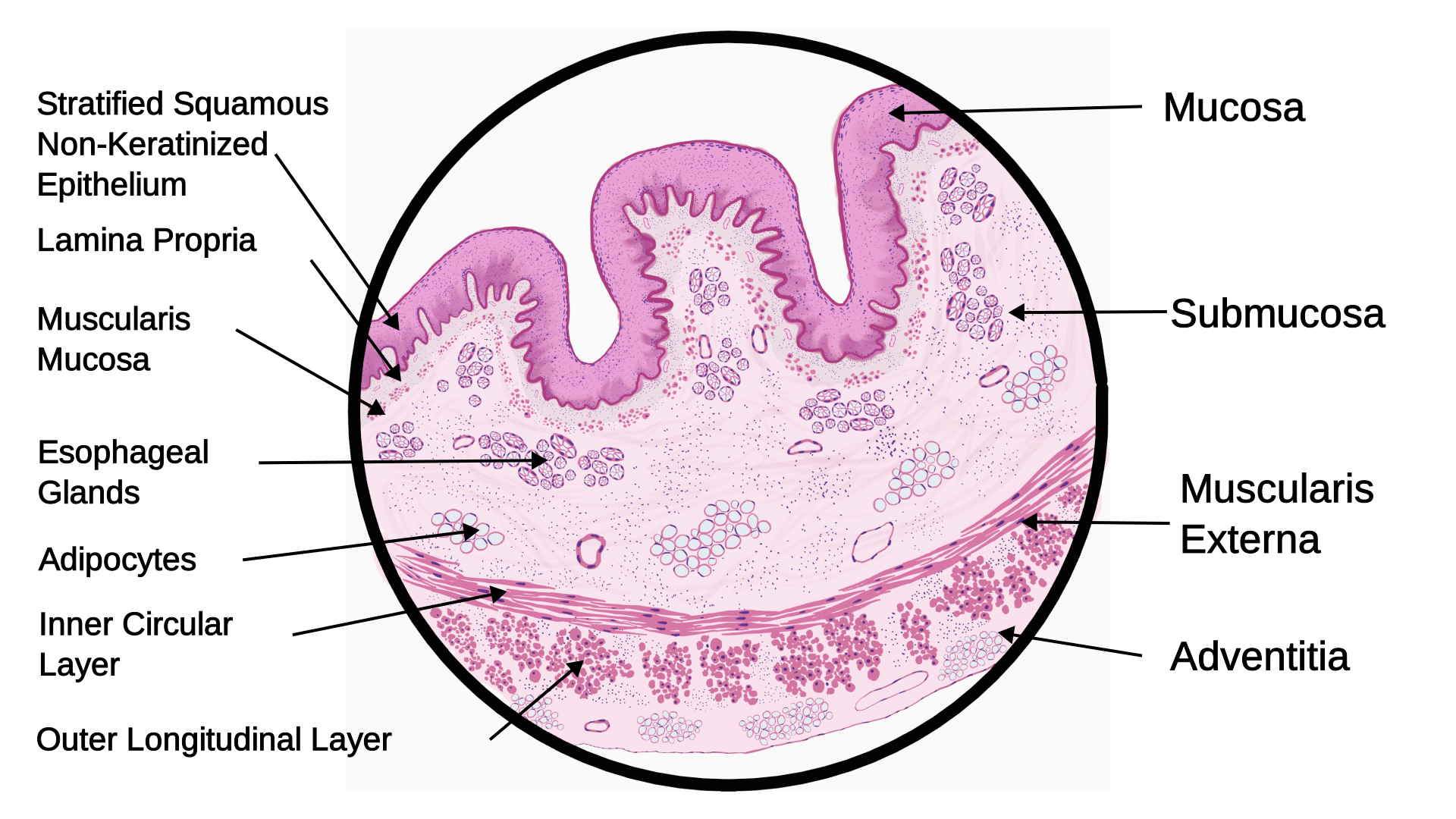
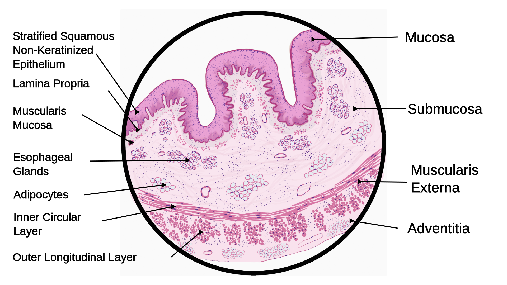
| Alternative text | Labelled histological cross-section of the oesophagus showing the mucosa with its stratified squamous non-keratinised epithelium, lamina propria and muscularis mucosa, the submucosa with oesophageal glands and adipocytes, the muscularis externa with inner circular and outer longitudinal layers, and the adventitia. |

Summary
| Description |
English: Schematic diagram of histology of Esophagus |
| Date | |
| Source | Own work |
| Author | DrJanaOfficial |
Licensing
I, the copyright holder of this work, hereby publish it under the following license:


This file is licensed under the Creative Commons Attribution 4.0 International license.
- You are free:
- to share – to copy, distribute and transmit the work
- to remix – to adapt the work
- Under the following conditions:
- attribution – You must give appropriate credit, provide a link to the license, and indicate if changes were made. You may do so in any reasonable manner, but not in any way that suggests the licensor endorses you or your use.
File history
Click on a date/time to view the file as it appeared at that time.
| Date/Time | Thumbnail | Dimensions | User | Comment | |
|---|---|---|---|---|---|
| current | 18:16, 7 June 2024 | | 1,920 × 1,080 (6.24 MB) | wikimediacommons>DrJanaOfficial | Uploaded own work with UploadWizard |
File usage
The following page uses this file: