
File:Merali 2019 neonatal ETT ultrasound simulator Fig1.jpg
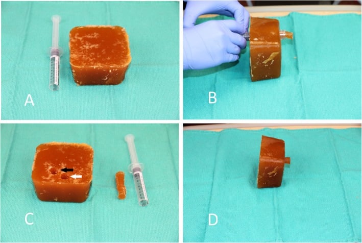
Low-cost ultrasound simulator for neonatal endotracheal tube placement identification. Four-panel construction photograph: (a) beef gelatine and psyllium fiber block with cut-off 10 mL syringe barrel, (b) syringe barrel creating staggered hollow cylinders within the block, (c) block with staggered hollow cylinders and retained plug from cylinder creation — superior cylinder is simulated trachea (white arrow), inferior cylinder is simulated esophagus (black arrow), (d) transverse anterior neck ultrasound image of the simulator showing simulated trachea (white arrow) and esophagus (black arrow).
Source: Merali HS, Tessaro MO, Ali KQ, Morris SK, Soofi SB, Ariff S (2019). "A novel training simulator for portable ultrasound identification of incorrect newborn endotracheal tube placement – observational diagnostic accuracy study protocol." BMC Pediatrics 19:434. DOI: 10.1186/s12887-019-1717-y. PMID: 31722685. PMC: PMC6852924.
Licence: CC BY 4.0 (BMC Pediatrics, BioMed Central / Springer Nature).
| Date created | Unknown |
|---|---|
| Author | Unknown |
| Date uploaded | April 18, 2026 |
| Uploader | Arturo Pelayo |
| License | Unknown |
File history
Click on a date/time to view the file as it appeared at that time.
| Date/Time | Thumbnail | Dimensions | User | Comment | |
|---|---|---|---|---|---|
| current | 23:25, 18 April 2026 | | 709 × 476 (90 KB) | Arturopelayo (talk | contribs) | TissueDB: Hero image for Qaim Ali Neonatal ETT Ultrasound Simulator — Merali 2019 Fig. 1, CC BY 4.0 |
You cannot overwrite this file.
File usage
The following page uses this file: