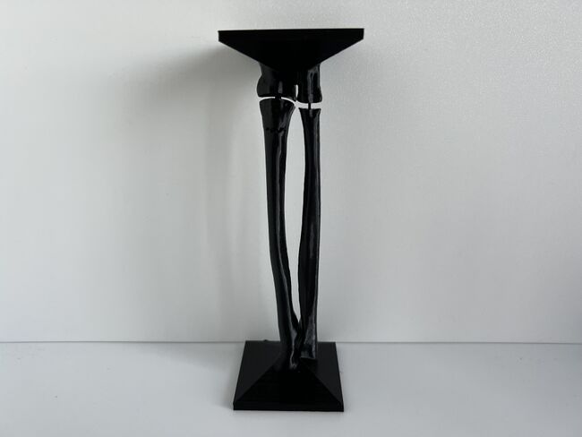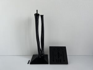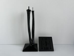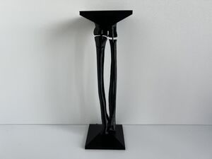
This module allows traditional bone setters, pre-hospital providers, clinical officers, nurses, nurse practitioners, and medical officers to become confident and competent in performing point-of-care ultrasound diagnostic imaging to rule out the presence of a pediatric distal forearm fracture and distinguish between buckle (torus) fractures and cortical break fractures to make appropriate referrals as part of the management of closed pediatric (< 16 years of age) distal forearm fractures in regions without access to X-ray imaging and orthopedic specialist coverage.[1][2][3][4][5][6][7][8][9] These 3D printed bone models feature a semi-engraved model number, gender symbol, and two mould insertion direction arrows on the base of each model to assist with model identification and proper orientation of each bone model in the mould.
3D Printed Pediatric Female Forearm Bone Models[edit | edit source]
In a 2022 Danish study on 4,316 pediatric patients, 98% of pediatric distal forearm fractures are isolated fractures and the incidence of distal forearm fractures in females was 44% and with a slighter higher incidence observed on left (57%) versus right (42%) extremities.[10] The incidence of pediatric distal forearm fractures peaks in girls between the ages of 9 to 11 years, with the highest incidence at age 10 years. The mean physeal plate width for the radius and ulna for girls ages 9-12 years of age was 0.26 and 0.25 cm, respectively.[11] Thus, a total radial length corresponding to the mean radial length of healthy Caucasian girls ages 10 years (185.39 mm) and a physeal width of 0.255 cm was used in the design of our gender-specific pediatric left forearm simulators.[11][12]
The fracture subtypes were selected based on input and X-rays provided by Dr. Peter J. Snelling, FACEM, FRACP (PEM & Gen Paeds), CCPU, Emergency Physician, Gold Coast University Hospital, Southport, Queensland, Australia, and Founder of Sonar Group (www.sonar.org.au), email: peter.j.snelling@gmail.com. A greenstick fracture with cortical breach of the palmar aspect of the distal radius and concurrent buckling of the dorsal radius will be included because it was the most commonly missed fracture by Australian nurse practitioners in a 2022 ultrasound training study.[13]
3D Printing Software, Hardware, and Filament Specifications[edit | edit source]
All 4 requirements below must be met in order to digitally manufacture the 3D Printed Pediatric Female Forearm Bone Models.
- 3D Printer Type: Fused Filament Fabrication
- Minimum Build Plate Z Height: 190 mm
- Filament: Unexpired Polylactic Acid (PLA) Fresh Out of the Sealed Packaging
- Filament Colour: White and Black PLA
3D Print White PLA Models for Unblinded Training[edit | edit source]
Download 3D Model STL Files For Slicing[edit | edit source]
Download the three 3D files (.STL) in each table below to input the required print settings to create the print files (.GCODE) for other 3D printers.
Table 1. No Fracture (Model #1 and #2)
| Model Number | Download STL File | Used Filament | Revision Date |
|---|---|---|---|
| 1 | Click on this link and click on "Model_1_-_Female_-29-Oct-2022_version.stl" to download the file | 39 grams | October 29, 2022 |
| 2 | Click on this link and click on "Model_2_-_Female_-29-Oct-2022_version.stl" to download the file | 25 grams | October 29, 2022 |
| Models 1 + 2 Connectors | Click on this link and click on "Models 1 + 2 Two_Connectors_-2-Nov-2022_version.stl" to download the file | 0.4 grams | November 2, 2022 |
Table 2. Buckle (Torus) Fracture of the Dorsal Distal Radius and Ulna (Model #3 and #4)
| Model Number | Download STL File | Used Filament | Revision Date |
|---|---|---|---|
| 3 | Click on this link and click on "Model_3_-_Female_-20-Feb-2023_version.stl" to download the file | 47 grams | February 20, 2023 |
| 4 | Click on this link and click on "Model_4_-_Female_-2-Nov-2022_version.stl" to download the file | 26 grams | November 2, 2022 |
| Models 3 + 4 Connectors | Click on this link and click on "Models 3 + 4 Two_Connectors_-2-Nov-2022_version.stl" to download the file | 0.4 grams | November 2, 2022 |
Table 3. Incomplete (Greenstick) Fracture of the Dorsal Distal Radius (Model #5 and #6)
| Model Number | Download STL File | Used Filament | Revision Date |
|---|---|---|---|
| 5 | Click on this link and click on "Model_5_-_Female_-3-Nov-2022_version.stl" to download the file | 44 grams | November 3, 2022 |
| 6 | Click on this link and click on "Model_6_-_Female_-5-Nov-2022_version.stl" to download the file | 32 grams | November 5, 2022 |
| Models 5 + 6 Connectors | Click on this link and click on "Models 5 + 6 Two_Connectors_-3-Nov-2022_version.stl" to download the file | 0.4 grams | November 3, 2022 |
Input Required Print Settings[edit | edit source]
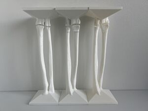
Please pay attention to and input all the required settings below to ensure the bone models are printed properly and display the required visual, tactile, and acoustic fidelity for orthopedic surgical simulation training.
- Slicing Program: use program designed for your 3D printer (i.e., use Prusa Slicer for a Prusa i3MK3S)
- Filament Material: **white PLA at original default settings** (if you have previously modified any values from the 3D slicer program's default settings for PLA filament, please change these values back to the default settings by clicking on the circular arrow to the left of each modified setting)
- Layer Height: 0.20 mm or less
- Infill: original default setting (not 0%)
- Support: None (click here for image)
- Build Plate Adhesion Type: no raft (click here for image)
- Model Orientation: flat surface (i.e., square base) of model lays flat on build plate
3D Print Black PLA Models for Blinded Training[edit | edit source]
Download 3D Model STL Files For Slicing[edit | edit source]
Download the 3D files (.STL) in the tables below to input the required print settings to create the print files (.GCODE) for other 3D printers.
Table 4. Incomplete (Greenstick) Fracture of the Dorsal Distal Radius (Model #5 and #6)
| Model Number | Download STL File | Used Filament | Revision Date |
|---|---|---|---|
| 5 | Click on this link and click on "Model_5_-_Female_-3-Nov-2022_version.stl" to download the file | 44 grams | November 3, 2022 |
| 6 | Click on this link and click on "Model_6_-_Female_-5-Nov-2022_version.stl" to download the file | 32 grams | November 5, 2022 |
| Models 5 + 6 Connectors | Click on this link and click on "Models 5 + 6 Two_Connectors_-3-Nov-2022_version.stl" to download the file | 0.4 grams | November 3, 2022 |
Table 5. Buckle (Torus) Fracture of the Volar Distal Radius (Model #7 and #8)
| Model Number | Download STL File | Used Filament | Revision Date |
|---|---|---|---|
| 7 | Click on this link and click on "Model_7_-_Female_-2-Nov-2022_version.stl" to download the file | 48 grams | November 2, 2022 |
| 8 | Click on this link and click on "Model_8_-_Female_-5-Nov-2022_version.stl" to download the file | 32 grams | November 5, 2022 |
| Models 7 + 8 Connectors | Click on this link and click on "Models 7 + 8 Two_Connectors_-2-Nov-2022_version.stl" to download the file | 0.2 grams | November 2, 2022 |
Table 6. Incomplete (Greenstick) Fracture of the Volar Distal Radius (Model #9 and #10)
| Model Number | Download STL File | Used Filament | Revision Date |
|---|---|---|---|
| 9 | Click on this link and click on "Model_9_-_Female_-3-Nov-2022_version.stl" to download the file | 45 grams | November 3, 2022 |
| 10 | Click on this link and click on "Model_10_-_Female_-5-Nov-2022_version.stl" to download the file | 32 grams | November 5, 2022 |
| Models 9 + 10 Connectors | Click on this link and click on "Models 9 + 10 Two_Connectors_-3-Nov-2022_version.stl" to download the file | 0.4 grams | November 3, 2022 |
Input Required Print Settings[edit | edit source]
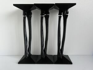
Please pay attention to and input all the required settings below to ensure the bone models are printed properly and display the required visual, tactile, and acoustic fidelity for orthopedic surgical simulation training.
- Slicing Program: use program designed for your 3D printer (i.e., use Prusa Slicer for a Prusa i3MK3S)
- Filament Material: **black PLA at original default settings** (if you have previously modified any values from the 3D slicer program's default settings for PLA filament, please change these values back to the default settings by clicking on the circular arrow to the left of each modified setting)
- Layer Height: 0.20 mm or less
- Infill: original default setting (not 0%)
- Support: None (click here for image)
- Build Plate Adhesion Type: no raft (click here for image)
- Model Orientation: flat surface (i.e., square base) of model lays flat on build plate
Assemble Pediatric Female Forearm Bone Models[edit | edit source]
Each pair of connectors is specifically designed for its corresponding numbered pair of bone models (i.e., only Models 1 + 2 Connectors should be inserted into Models 1 + 2).
Align the female part of the radius of the distal forearm over the male connector of the radius of the proximal forearm to connect the radius of the distal forearm to the proximal forearm. Align the female part of the ulna of the distal forearm over the male connector of the ulna of the proximal forearm to connect the ulna of the distal forearm to the proximal forearm.
Acknowledgements[edit | edit source]
This work is funded by a grant from the Intuitive Foundation. Any research, findings, conclusions, or recommendations expressed in this work are those of the author(s), and not of the Intuitive Foundation.
References[edit | edit source]
- ↑ Onyemaechi NO, Itanyi IU, Ossai PO, Ezeanolue EE. Can traditional bonesetters become trained technicians? Feasibility study among a cohort of Nigerian traditional bonesetters. Hum Resour Health. 2020 Mar 20;18(1):24. doi: 10.1186/s12960-020-00468-w. PMID: 32197617; PMCID: PMC7085192.
- ↑ Heiner JD, McArthur TJ. The ultrasound identification of simulated long bone fractures by prehospital providers. Wilderness Environ Med. 2010 Jun;21(2):137-40. doi: 10.1016/j.wem.2009.12.028. Epub 2009 Dec 22. PMID: 20591377.
- ↑ Heiner JD, Baker BL, McArthur TJ. The ultrasound detection of simulated long bone fractures by U.S. Army Special Forces Medics. J Spec Oper Med. 2010 Spring;10(2):7-10. PMID: 20936597.
- ↑ Heiner JD, Proffitt AM, McArthur TJ. The ability of emergency nurses to detect simulated long bone fractures with portable ultrasound. Int Emerg Nurs. 2011 Jul;19(3):120-4. doi: 10.1016/j.ienj.2010.08.004. Epub 2010 Sep 25. PMID: 21665155.
- ↑ Snelling PJ, Jones P, Keijzers G, Bade D, Herd DW, Ware RS. Nurse practitioner administered point-of-care ultrasound compared with X-ray for children with clinically non-angulated distal forearm fractures in the ED: a diagnostic study. Emerg Med J. 2021 Feb;38(2):139-145. doi: 10.1136/emermed-2020-209689. Epub 2020 Sep 8. PMID: 32900856.
- ↑ Snelling PJ, Jones P, Moore M, Gimpel P, Rogers R, Liew K, Ware RS, Keijzers G. Describing the learning curve of novices for the diagnosis of paediatric distal forearm fractures using point-of-care ultrasound. Australas J Ultrasound Med. 2022 Mar 7;25(2):66-73. doi: 10.1002/ajum.12291. PMID: 35722050; PMCID: PMC9201201.
- ↑ Heiner JD, McArthur TJ. A simulation model for the ultrasound diagnosis of long-bone fractures. Simul Healthc. 2009 Winter;4(4):228-31. doi: 10.1097/SIH.0b013e3181b1a8d0. PMID: 19915442.
- ↑ Snelling PJ, Keijzers G, Byrnes J, Bade D, George S, Moore M, Jones P, Davison M, Roan R, Ware RS. Bedside Ultrasound Conducted in Kids with distal upper Limb fractures in the Emergency Department (BUCKLED): a protocol for an open-label non-inferiority diagnostic randomised controlled trial. Trials. 2021 Apr 14;22(1):282. doi: 10.1186/s13063-021-05239-z. PMID: 33853650; PMCID: PMC8048294.
- ↑ Snelling PJ. A low-cost ultrasound model for simulation of paediatric distal forearm fractures. Australas J Ultrasound Med. 2018 Feb 25;21(2):70-74. doi: 10.1002/ajum.12083. PMID: 34760505; PMCID: PMC8409885.
- ↑ Korup LR, Larsen P, Nanthan KR, Arildsen M, Warming N, Sørensen S, Rahbek O, Elsoe R. Children's distal forearm fractures: a population-based epidemiology study of 4,316 fractures. Bone Jt Open. 2022 Jun;3(6):448-454. doi: 10.1302/2633-1462.36.BJO-2022-0040.R1. PMID: 35658607; PMCID: PMC9233428.
- ↑ 11.0 11.1 Ng L, Saul T, Lewiss RE. Sonographic baseline physeal plate width measurements in healthy, uninjured children. Pediatr Emerg Care. 2014 Dec;30(12):871-4. doi: 10.1097/PEC.0000000000000290. PMID: 25407037.
- ↑ Gindhart PS. Growth standards for the tibia and radius in children aged one month through eighteen years. Am J Phys Anthop 1973; 39(1): 41–8.
- ↑ Snelling PJ, Jones P, Moore M, Gimpel P, Rogers R, Liew K, Ware RS, Keijzers G. Describing the learning curve of novices for the diagnosis of paediatric distal forearm fractures using point-of-care ultrasound. Australas J Ultrasound Med. 2022 Mar 7;25(2):66-73. doi: 10.1002/ajum.12291. PMID: 35722050; PMCID: PMC9201201.
