Aortic Annulus, Aortic Root, and Aorta
George Gellert, Aortic annulus anatomy and measurement and its relevance to successful transcatheter aortic valve replacement. [cited 2020 Sep 14].
- Aortic valve structure and shape (irregular shaped oval or elliptical)
- Explains aortic root anatomy
- Basal ring measurement method
Blanke P, Siepe M, Reinöhl J, Zehender M, Beyersdorf F, Schlensak C, et al. Assessment of aortic annulus dimensions for Edwards SAPIEN Transapical Heart Valve implantation by computed tomography: calculating average diameter using a virtual ring method. European Journal of Cardio-Thoracic Surgery. 2010 Dec [cited 2020 Sep 14];38(6):750–8.
- Study population: 71 patients with severe symptomatic aortic stenosis
- Aortic annulus dimensions in coronal and sagittal view
Evangelista A, Flachskampf FA, Erbel R, Antonini-Canterin F, Vlachopoulos C, Rocchi G, et al. Echocardiography in aortic diseases: EAE recommendations for clinical practice. European Journal of Echocardiography. 2010 Sep 1 [cited 2020 Sep 14];11(8):645–58. DOI:10.1093/ejechocard/jeq056
- Dimensions of the aortic annulus, ascending aorta, aortic arch, and descending aorta
Suchá D, Tuncay V, Prakken NHJ, Leiner T, van Ooijen PMA, Oudkerk M, et al. Does the aortic annulus undergo conformational change throughout the cardiac cycle? A systematic review. European Heart Journal – Cardiovascular Imaging. 2015 Sep 15 [cited 2020 Sep 14];jev210.
- Aortic annulus dimension from 29 articles
- 7 studies of a healthy population, 10 studies comparing healthy population with aortic stenosis, and 12 studies of aortic stenosis population
- Total of 2021 patients
Zhu D, Zhao Q. Dynamic Normal Aortic Root Diameters: Implications for Aortic Root Reconstruction. The Annals of Thoracic Surgery. 2011 Feb [cited 2020 Sep 14];91(2):485–9.
- Study population: 314 healthy adults age ranging from 17 to 60 years
- Dimensions of aortic annulus and aortic root
Son MK, Chang S-A, Kwak JH, Lim HJ, Park S-J, Choi J-O, et al. Comparative measurement of aortic root by transthoracic echocardiography in normal Korean population based on two different guidelines. Cardiovascular Ultrasound [Internet]. 2013 Aug 13 [cited 2020 Sep 14];11(1).
*Study population: 112 Korean adults age ranging 20-69 years with no cardiovascular disease
- Dimensions of aortic annulus and aortic root
Boufi M, Guivier-Curien C, Loundou AD, Deplano V, Boiron O, Chaumoitre K, et al. Morphological Analysis of Healthy Aortic Arch. European Journal of Vascular and Endovascular Surgery [Internet]. 2017 May [cited 2020 Sep 14];53(5):663–70.
- Study population: 123 healthy population with no history of cardiovascular disease
- Shows different aortic arch segments and their dimensions
Mitral Valve
Naoum C, Leipsic J, Cheung A, Ye J, Bilbey N, Mak G, et al. Mitral Annular Dimensions and Geometry in Patients With Functional Mitral Regurgitation and Mitral Valve Prolapse. JACC: Cardiovascular Imaging. 2016 Mar [cited 2020 Sep 14];9(3):269–80. DOI: 10.1016/j.jcmg.2015.08.022
- D-shaped mitral annulus concept
- A study performed on 88 control group patients, 32 mitral valve prolapse, and 27 functional mitral regurgitation
- Dimensions of mitral valve annulus (area, perimeter, trigone to trigone, septal to lateral, and intercommissural)
Abdelghani M, Spitzer E, Soliman OII, Beitzke D, Laggner R, Cavalcante R, et al. A simplified and reproducible method to size the mitral annulus: implications for transcatheter mitral valve replacement. European Heart Journal – Cardiovascular Imaging. 2016 Jun 26 [cited 2020 Sep 14];jew132.
- D shaped and saddle shape mitral annulus model comparison
- Study conducted on 47 patients who had done CRA for coronary revascularization or transcatheter aortic valve replacement
- Three mitral annular diameter methods: Dmean, Darea, and Dperimeter
- Dimensions of mitral annular height
Left Ventricle
Ho SY. Anatomy and myoarchitecture of the left ventricular wall in normal and in disease. European Journal of Echocardiography [Internet]. 2009 Nov 4 [cited 2020 Sep 14];10(8):iii3–7.
- Explains anatomy of the left ventricle
Chetrit M, Roujol S, Picard MH, Timmins L, Manning WJ, Rudski LG, et al. Optimal Technique for Measurement of Linear Left Ventricular Dimensions. Journal of the American Society of Echocardiography [Internet]. 2019 Apr [cited 2020 Sep 14];32(4):476-483.e1.
- Study population: Derivation study-75 patients, Validation study-100 patients
- Left ventricular dimensions at the midventricular level

 Left Ventricle
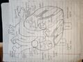 Full Heart
