Video Summary[edit | edit source]
Osteotomy planning[edit | edit source]
Bone segments and axes[edit | edit source]

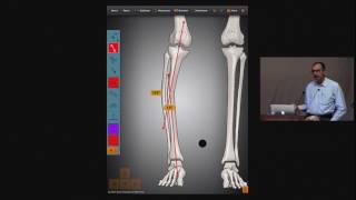
When there is Malalignment or Malorientation in femur or tibia bones, the bone with deformity can be divided into proximal and distal bone segments. The proximal and distal bone segments have their own proximal mechanical axis (PMA) or proximal anatomical axis (PAA) line and distal mechanical axis (DMA) or distal anatomical axis (DAA) line respectively (Fig. 8a)
Center of rotation of angulation[edit | edit source]

The point at which the proximal and distal axis lines intersect is known as the Center of rotation of angulation (CORA). (Fig. 8b)
The PMA or DMA lines can be drawn from respective joint centers taking normal values of joint orientation angle as reference (Fig. 8b).
The acute angle between the two axis lines (PMA and DMA) or (PAA and DAA) is the 'Magnitude (Mag) of correction' required to correct the deformity in the bone (Fig. 8b). This is also called the 'Correction angle'.
Angulation correction axis[edit | edit source]

The PAA and DAA lines can be drawn taking mid-diaphyseal lines in the proximal shaft or the distal shaft bone segment respectively or drawing axis lines taking normal values of anatomical angles from the joint lines (Fig. 8c).
The bone deformity can be corrected by performing an osteotomy (bone cut) near CORA calculated from the deformity planning in frontal plane and rotating the distal bone segment by 'Correction angle' about an imaginary Angulation Correction Axis (ACA) which may or may not pass through CORA (Fig. 8b). Many times ACA is also referred to as 'hinge' or 'pivot' about which the resected bone segment rotates.
Rules of bone osteotomy[edit | edit source]
There are three rules of Osteotomy which summarise the correction of a deformed bone.
First rule of osteotomy[edit | edit source]

If the ACA passes through the CORA (ACA-CORA), and the osteotomy line or level also passes through CORA, then the resected bone segment will only angulate with respect to the other bone segment without any translation or displacement at the osteotomy level. The proximal axis and distal axis lines will become collinear and parallel if the magnitude of angulation is equal to the 'Correction angle' (Fig. 8d)
Second rule of osteotomy[edit | edit source]

If the ACA passes through the CORA (ACA-CORA), but the osteotomy line or level is above or below the CORA, then the resected bone segment will angulate and also translate with respect to the other bone segment at the osteotomy level. The proximal axis and distal axis lines will become collinear and parallel if the magnitude of angulation is equal to 'Correction angle' (Fig. 8e)
Third rule of osteotomy[edit | edit source]

If the ACA does not pass through the CORA, and the osteotomy line or level is at the ACA, then the resected bone segment will only angulate with respect to the other bone segment at the osteotomy level. The proximal axis and distal axis lines will become only parallel and not collinear, if the magnitude of angulation is equal to 'Correction angle'. The resulting bone correction will induce a 'translation deformity' between the two bone segments. (Fig. 8f)
Types of osteotomy[edit | edit source]
The osteotomy can be performed by doing an opening wedge osteotomy, a closing wedge osteotomy or a neutral wedge osteotomy.
Opening wedge osteotomy[edit | edit source]

In this kind of osteotomy, the ACA-CORA is at the convex cortex of the bone and also called as the opening wedge point. The osteotomy line or level passes through the opening wedge point. The convex cortex remains in contact whereas the concave side is fully distracted or opened by an angle equal to the correction angle in angular terms and a length 'L' at the most concave side. The bone is lengthened by the same length 'L' (Fig. 8g)
Closed wedge osteotomy[edit | edit source]

Closing wedge osteotomy: In this kind of osteotomy, the ACA-CORA is at the concave cortex of the bone and also called as the closing wedge point.
The osteotomy line or level passes through the closing wedge point. The concave cortex remains in contact whereas at the convex side, the bone wedge is removed by an angle equal to the correction angle in angular terms and a length 'L' at the most convex side. The bone is shortened by the same length 'L'.
The two bone segments come in full bone-to-bone contact at the end of correction (Fig. 8h)
Neutral wedge osteotomy[edit | edit source]

Neutral wedge osteotomy: In this kind of osteotomy, the ACA-CORA is in between the concave and convex cortex of the bone and also called as the neutral wedge point. The osteotomy line or level passes through the neutral wedge point. There is partial opening wedge at the concave cortex and partial closing wedge at the convex cortex. The bone wedge is removed by an angle equal to the correction angle in angular terms and a length 'L/2' at the most convex side compared to the closing wedge osteotomy length 'L'. The bone is lengthened by the length 'L/2' compared to the opening wedge length 'L'.
All parts of the bone convex to the neutral wedge point come in full bone-to-bone contact whereas in the concave part reactive to the neutral wedge there is a gap between the bone segments (Fig. 8i)