(33 intermediate revisions by 4 users not shown)
Line 1: Line 1:
{{MOST}}
{{MOST}}
{{MY5970}}


[[category: MOST methods]]
[[category: MOST methods]]


==Introduction==
==Introduction==
*basic theory - heavily referenced (focus primarily on ingan PL) after initial theory of concept
Photoluminescence{{w|Photoluminescence}} is the process where a substance absorbs photons and re-emits photons.  The incident photons excite electrons from a lower energy level to a higher energy level.  As the electrons relax from the higher energy level to the lower energy level a photon is released.  For a [[semiconductor]], the electrons are excited from a set of energy levels called the [[valence band]] to a higher set of energy levels called the [[conduction band]].  The energy area between the valence band and the conduction band is called the [[band gap]], and no electrons can be energetically found here.


== Design ==
'''Photoluminescence (PL) Spectroscopy''' is a type of contactless and nondestructive probing method for determining the electronic structure of a sample.  In PL Spectroscopy, a [[laser]] is fired at a sample and the resulting fluorescence is measured by a [[spectrometer]].  The measured fluorescence can be used to determine the band gap of a semiconductor sample, impurity levels and possible defects in the sample, and the recombination mechanisms within the sample [http://www.nrel.gov/pv/measurements/photoluminescence_spectroscopy.html]. The determination of the electronic band gap for a semiconductor is the primary focus of this work.
*equipment list and specs for our ocean optics system
*Design schematic
*pictures of setup
* operation instructions
  this section will eventually get ported to its own protocol page.


== Open source PL Design ==
As previously stated, in a semiconductor the electrons are excited from the valence to the conduction band.  When electrons relax from the conduction band back down to the valence band, photons will be released.  '''These photons will have energy that matches the transition from the conduction band to the valence band.  Measuring the emitted photons’ wavelength with a spectrometer will reveal the energy of the photon and therefore the size of the bandgap.'''
*equipment list and specs
*3D design schematic - particularly of external case to be printed by reprap
*pictures of setup
* operation instructions


== See also ==
== Design ==
=== Equipment List ===
*Pavilion Integration Corp W445-40FS 445 nm laser - 500mW
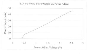
[[image:ZIC 002.jpg|thumb|Laser linear response]]
*Thor Labs LB1761-A N-BK7 Bi-Convex Lens, Ø1", f = 25.4 mm, Anit-Reflective Coating: 350-700nm
*Thor Labs KM100-E02 Kinematic Mount for Ø1" Optics with Visible Laser Quality Mirror
*Ocean Optics USB2000+VIS-NIR spectrometer, NIRQuest 512-2.5
*Ocean Optics 600 micron VIS NIR fiber optics cable part # QP600-2-VIS-NIR
*[[open-source_optics_base|Open-Source Optics Base]]
*[[open-source_simple_semiconductor_sample_holder|Open-Source Simple Semiconductor Sample]]
*[[open-source_lens_holder|Open-Source Lens Holder]]
*[[open-source_mirror_mount|Open-Source Mirror Mount]]
*[[open-source_fiber_optic_cable_holder|Open-Source Fiber Optic Cable]]


* [[Photoluminescence protocol:QAS]]
===Design Schematic===
* [[PL data curve fitting:QAS]]
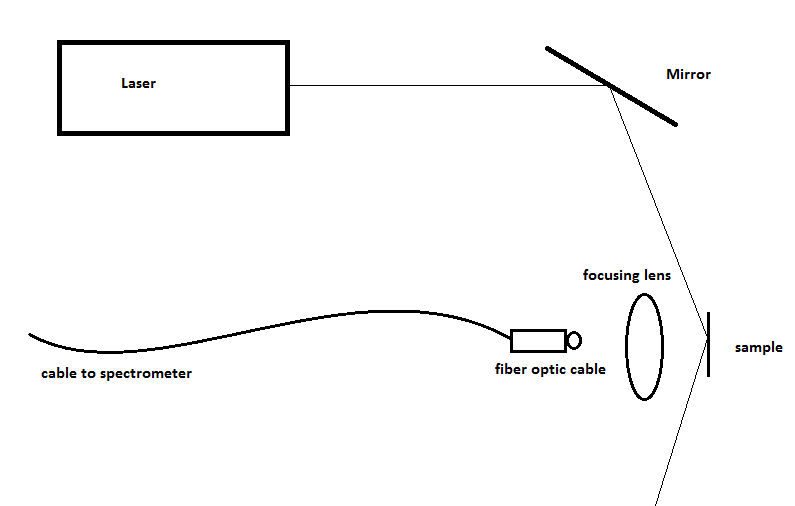
[[Image:Open-source-PL_setup.png]]
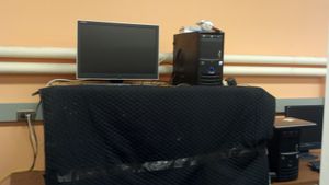
[[Image:PL-test-bench.jpg|thumb|left|Fig 1. The PL Test Bench]]
[[Image:PL-setup.jpg|thumb|left|Fig 2. The PL Setup]]


== Literature Review ==
===Design Considerations and Explanations===
The first step to using of PL spectroscopy is to have a powerful enough laser to excite the electrons from the valence band to the conduction band for all the different samples you wish to examine.  We used at 450 nm (2.755 eV) laser to test samples.  This will only work for semiconductors with bandgaps smaller than 2.755eV. When choosing a laser, it is the wavelength that is important with intensity being a secondary concern since it is the wavelength that determines if the electrons jump the band gap.


  This section is currently being updated...
The setup of the PL system was designed from two separate works from the University of Colorado [http://www.physics.ohio-state.edu/~reu/99reu/final_reports/paper_hall.PDF] and from the University of Ohio [http://www.physics.ohio-state.edu/~reu/99reu/final_reports/paper_hall.PDF].  While researching the design of a PL system, it became obvious that researchers in general do not share their exact PL system design. Both of the works feature explicit diagrams of their photoluminescence spectroscopy setups. This open source PL spectroscopy setup is largely based on their previous work.


=== Papers/Documents ===
Both of the setups from Colorado and OSU used a neutral density filter. We do not use one here because our laser has a variable intensity dial on it. The use of a mirror to bounce the laser onto the sample instead of direct illumination from the laser offers more flexibility. It is much easier to adjust the mirror than the laser. The laser is incident on the sample at a glancing angle so that specular reflection from the sample does not go near the fiber optic cable. This will reduce the laser signal read on the spectrometer and thus the noise. The spectrometer will still read some of the laser signal from diffuse reflection. The focusing lens is used to gather the fluorescence from the sample and focus it onto the fiber optic cable. Thus the fiber optic cable must be placed away from the lens at a distance equal to the focal length of the lens. The whole PL experiment is done within a dark box to limit the effect of outside light on the spectrometer.
====[http://ieeexplore.ieee.org/xpl/freeabs_all.jsp?arnumber=299204 A Low Cost Compact CCD Grating Spectrometer]<ref> K. F. Lin and G. Schaefer, “A low cost compact CCD grating spectrometer,” in , Conference Record of the 1993 IEEE Industry Applications Society Annual Meeting, 1993, 1993, pp. 2334–2337 vol.3.</ref>====
''Abstract''<br />
The authors describe a grating spectrometer consisting of a slit, a transmission holographic grating, a lens, and a CCD (charge coupled device) camera. A frame grabber equipped in a computer digitizes the NTSC signal from the CCD camera. The system is relatively small (12.7 cm×17.8 cm×30.5 cm) and light-weight (5.4 kg). The system will measure the output of modern fluorescent or compact fluorescent lamps more accurately than conventional filter-based photometers.


====[http://ieeexplore.ieee.org/xpl/freeabs_all.jsp?arnumber=1277106 Tolerance design of optical micro-bench by statistical design of experiment]<ref>
The mirror, sample, lens, and fiber optic cable are held in place with [[open source optics]] holders, which can be printed on an [[open source 3D printer]]. These open source optic holders allow us greater flexibility of placement than an optical bread board or lab bench would. However, it does lower the overall stability of the system, but such fined tuned stability is not needed for a simple PL system like this.
B. C. Hwang, H. Y. Park, J. Y. Lee, S. G. Park, S. G. Lee, B. H. O, D. S. Choi, and E. H. Lee, “Tolerance design of optical micro-bench by statistical design of experiment,” in CLEO/Pacific Rim 2003 - The 5th Pacific Rim Conference on Lasers and Electro-Optics, 2003, vol. 2.
</ref>====
''Abstract''<br />
Design rule of optical micro-bench and tolerance of positional error of components assembled onto the bench are investigated in case of simple linear connections of incoming and outgoing optical fibers with and without ball lenses. Since there are many error variables with wide range of values that affects the optical coupling, the efficient array with reduced number of combinations is generated using statistical design of experiment and coupling efficiencies are calculated. From 3d B point, the tolerance of each position error is determined. For the fiber-to-fiber connection with ball lenses, the longitudinal and lateral positional errors have the strong interaction with each other to the coupling efficiency and thus should be be limited simultaneously.


====[http://rsi.aip.org/resource/1/rsinak/v77/i2/p023907_s1?isAuthorized=no Laser photoluminescence spectrometer based on charge-coupled device detection]<ref> O. H. Y. Zalloum, M. Flynn, T. Roschuk, J. Wojcik, E. Irving, and P. Mascher, “Laser photoluminescence spectrometer based on charge-coupled device detection for silicon-based photonics,” Review of Scientific Instruments, vol. 77, no. 2, p. 023907–023907–8, Feb. 2006.
===Improvements===
</ref>====
There many improvements to this simple system that could be implemented.  
''Abstract''<br />
We describe and characterize a multichannel modular room temperature photoluminescence spectroscopy system. This low cost instrument offers minimization of size and complexity as well as good flexibility and acceptable spectral resolution. The system employs an efficient flexible front end optics and a sensitive spectrometer with a charge-coupled device array detector. The spectrometer has no moving parts and is more robust than a scanning system. The scientific motivation was to enable the photoluminescence study of various silicon photonics structures. Typical applications are presented for SiOx (x<2) films. It is demonstrated that high-quality steady state photoluminescence data with excellent signal to noise enhancement capability can be delivered besides the ability to perform simultaneous multiwavelength measurements in one shot. This instrument is shown to be useful for evaluating semiconductor wafers, including those intended for light emitting structures from silicon-based photonic crystals. The design, construction, calibration, and the unique features of this system are presented, and performance tests of a prototype are discussed.


====[http://www.springerlink.com/content/hrv6843k18517660/ The Spectral Irradiance Monitor: Scientific Requirements, Instrument Design, and Operation Modes]<ref>
[[image:Sampleh3.jpg|thumb|left|Fig. 3. Open-source sample holder v3]]  
J. Harder, G. Lawrence, J. Fontenla, G. Rottman, and T. Woods, “The Spectral Irradiance Monitor: Scientific Requirements, Instrument Design, and Operation Modes,” in The Solar Radiation and Climate Experiment (SORCE), G. Rottman, T. Woods, and V. George, Eds. New York, NY: Springer New York, pp. 141–167. </ref>====
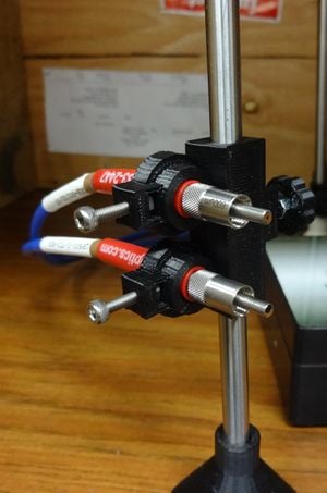
[[image:Switcher.jpg|thumb|right|Fig. 4. Open-source fiber switcher]]
''Abstract''<br />
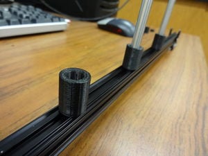
[[image:Rail.JPG|thumb|left|Fig. 5. Open-source rail]]
The Spectral Irradiance Monitor (SIM) is a dual Fèry prism spectrometer that employs 5 detectors per spectrometer channel to cover the wavelength range from 200 to 2700 nm. This instrument is used to monitor solar spectral variability throughout this wavelength region. Two identical, mirror-image, channels are used for redundancy and in-flight measurement of prism degradation. The primary detector for this instrument is an electrical substitution radiometer (ESR) designed to measure power levels ∼1000 times smaller than other radiometers used to measure TSI. The four complementary focal plane photodiodes are used in a fast-scan mode to acquire the solar spectrum, and the ESR calibrates their radiant sensitivity. Wavelength control is achieved by using a closed loop servo system that employs a linear charge coupled device (CCD) in the focal plane. This achieves 0.67 arcsec control of the prism rotation angle; this is equivalent to a wavelength positioning error of δλ/λ = 150 parts per million (ppm). This paper will describe the scientific measurement requirements used for instrument design and implementation, instrument performance, and the in-flight instrument operation modes.


====[http://lib.semi.ac.cn:8080/tsh/dzzy/wsqk/SPIE/vol4721/4721-26.pdf Indium Gallium Arsenide Imaging with Smaller Cameras, Higher Resolution Arrays, and Greater Material Sensitivity] <ref>Martin H. Ettenberg, Marshall J. Cohen, Robert M. Brubaker, Michael J. Lange, Matthew T. O’Grady, and Gregory H. Olsen, “Indium Gallium Arsenide Imaging with Smaller Cameras, Higher Resolution Arrays, and Greater Material Sensitivity.” SPIE, vol. 4721, Proc. of SPIE, p. 4721–26, 2002. </ref>====
* Shown on the left is an improved open-source sample holder for pieces of semiconductor wafers . Instead of the clamping a sample, it provides a 2mm lip that allows samples to fluently sink into the slot. The purpose of the design is to make sample switching faster and easier, and reduce abrasion caused by frequent sample switch. Also notice the hemisphere hole in the backside makes it easier for tweezers work. The design can be applied to variety of wafer-like material, we also use it as a mirror holder since it works fine with our appropediately sized mirror.
''Abstract''<br />
Indium Gallium Arsenide (InGaAs) photodiode arrays have numerous commercial, industrial, and military applications. During the past 10 years, great strides have been made in the development of these devices starting with simple 256-element linear photodiode arrays and progressing to the large 640 x 512 element area arrays now readily available. Linear arrays are offered with 512 elements on a 25 micron pitch with no defective pixels, and are used in spectroscopic monitors for wavelength division multiplexing (WDM) systems as well as in machine vision applications. A 320 x 240 solid-state array operates at room temperature, which allows development of a camera which is smaller than 25 cm<sup>3</sup> in volume, weighs less than 100 g and uses less than 750 mW of power. Two dimensional focal plane arrays and cameras have been manufactured with detectivity, D*, greater than 1014 cm-√Hz/W at room temperature and have demonstrated the ability to image at night. Cameras are also critical tools for the assembly and performance monitoring of optical switches and add-drop multiplexers in the telecommunications industry. These same cameras are used for the inspection of silicon wafers and fine art, laser beam profiling, and metals manufacturing. By varying the Indium content, InGaAs photodiode arrays can be tailored to cover the entire short-wave infrared spectrum from 1.0 micron to 2.5 microns. InGaAs focal plane arrays and cameras sensitive to 2.0 micron wavelength light are now available in 320 x 240 formats.


==== [http://ieeexplore.ieee.org/xpl/freeabs_all.jsp?arnumber=1426423 A Compact Spectrometer based on a Micromachined Torsional Mirror Device]<ref>
* On the right is the improved fiber holder is a dual-channel fiber switcher, which attaches two fiber optic cable to a 8mm smooth rod and allow easy switching between cables for optical experiments.
A. Kenda, W. Scherf, R. Hauser, H. Gruger, and H. Schenk, “A compact spectrometer based on a micromachined torsional mirror device,” in Proceedings of IEEE Sensors, 2004, 2004, pp. 1312– 1315 vol.3.
</ref> ====
''Abstract''<br />
We describe a spectrometer employing a microelectromechanical system (MEMS). A torsional micromirror device, which was originally designed for laser scanner applications, has been modified to act as a diffraction grating in a Czerny-Turner setup. This spectrometer system combines the working principle of a scanning monochromator with the compactness of a microsystem. The micromirror device is operated in a harmonic scanning mode at a resonant frequency of 500 Hz, enabling the system to acquire a spectrum per half cycle in one millisecond. The spectra of a calibration source in the visible and near-infrared spectral range as well as simulations concerning the optical system demonstrate the performance of the spectrometer.


====[http://ieeexplore.ieee.org/xpl/freeabs_all.jsp?arnumber=5164008 Surface Photovoltage Spectroscopy—A New Approach to the Study of High-Gap Semiconductor Surfaces]<ref>H. C. Gatos and J. Lagowski, “Surface Photovoltage Spectroscopy—A New Approach to the Study of High-Gap Semiconductor Surfaces,” Journal of Vacuum Science and Technology, vol. 10, no. 1, pp. 130–135, Jan. 1973.
* In order to smoothly move each parts in our PL system, we've designed an optical rail that helps accurately align our system in a line and make lens focusing much easier. Installed with strong magnet the rail is clinging to the platform firmly. Now the system works in a fixed X-Y plane with measurable precision.
</ref> ====
''Abstract''<br />
Surface photovoltage spectroscopy is based on the photostimulated depopulation and population of surface states brought about by sub-bandgap monochromatic illumination, while the over-all number of bulk free carriers remains essentially unchanged. Such transitions and their transients (as determined by changes is the contact potential difference) allow the direct determination of the energy positions and the dynamic parameters of the surface states. Surface photovoltage spectroscopy was successfully applied to the surfaces of CdS, ZnO, and GaAs. A model was developed which accounts for the processes involved in surface photovoltage spectroscopy.


====[http://books.google.com/books?id=dNx1OLgn1xcC Laser Spectroscopy: Basic Concepts and Instrumentation]<ref>W. Demtröder, Laser spectroscopy: basic concepts and instrumentation. Springer, 2003.
==== Further improvement ====
</ref>====
* A component for a low temperature liquid to cool the sample during data taking.
''Abstract''<br />
* The addition of a optical chopper and lock-in amplifier would improve signal to noise ratio
Keeping abreast of the latest techniques and applications on laser spectroscopy. While the general concept is unchanged, new features a broad array of new material, e.g., frequency doubling in external cavities, reliable cw-parametric oscillators, tunable narrow-band UV sources, more sensitive detection techniques, tunable femtosecond and sub-femtosecond lasers (X-ray region and the attosecond range), control of atomic and molecular excitations, frequency combs able to synchronize independent femtosecond lasers, coherent matter waves, and still more applications in chemical analysis, medical diagnostics, and engineering.
* A monochromator would allow the system to focus on a small range of wavelengths at a time instead of looking a wide range of wavelengths.


==== XX ====
As part of the open source optics project we will be designing all of these components.


=== Patents/IP ===
== To operate ==
====[http://patft.uspto.gov/netacgi/nph-Parser?Sect2=PTO1&Sect2=HITOFF&p=1&u=/netahtml/PTO/search-bool.html&r=1&f=G&l=50&d=PALL&RefSrch=yes&Query=PN/8040507 Spectrometer - #8,040,707]<ref> K. Shibayama, “United States Patent: 8040507 - Spectrometer,” U.S. Patent 8040507 18-Oct-2011.</ref>====
* [[Spectrometer and light source calibration: MOST]]
''Abstract''<br />
In the spectrometer 1, a lens portion 3 having a spherical surface 35 on which a spectroscopic portion 4 is provided and a bottom plane 31 in which a light detecting element 5 is disposed, has a side plane 32 substantially perpendicular to the bottom plane 31 and a side plane 34 substantially perpendicular to the bottom plane 31 and the side plane 32. Then, a package 11 that houses a spectroscopy module 10 has side planes 16 and 18 respectively coming into planar-contact with the side planes 32 and 34, and contact portions 22 coming into contact with the spherical surface 35. Therefore, the side planes 32 and 34 of the lens portion 3 are respectively brought into planar-contact with the side planes 16 and 18 of the package 11 while bringing the spherical surface 35 of the lens portion 3 into contact with the contact portions 22 of the package 11, that positions the spectroscopic portion 4 and the light detecting element 5 with respect to a light incident window plate 25 of the package 11.


====[http://patft.uspto.gov/netacgi/nph-Parser?Sect1=PTO1&Sect2=HITOFF&d=PALL&p=1&u=%2Fnetahtml%2FPTO%2Fsrchnum.htm&r=1&f=G&l=50&s1=7,437,000.PN.&OS=PN/7,437,000&RS=PN/7,437,000 Full Spectrum Detecting Pixel Camera - #7,437,000]<ref>
[[category:open source optics]]
E. Rosenthal, R. J. Solomon, and C. Johnson, “United States Patent: 7437000 - Full spectrum color detecting pixel camera,” U.S. Patent 743700014-Oct-2008.
</ref>====
''Abstract''<br />
This invention comprises the means for the capture of full spectrum images in an electronic camera without the use of color primary filters to limit the spectral color gamut of the captured image. The fundamental principle of the invention is that each pixel of the image sensor acts as an independent spectrophotometer and spectral separator. Electromagnetic energy enters though a slit or collimating optic. Electromagnetic energy gets diffracted into component spectra by diffraction grating spectrophotometer for each pixel of image Electromagnetic energy leaves diffraction grating at different angles based on wavelength of the energy Spectrophotometer separates light for each pixel into its spectral components onto photodetector line array elements. Individual line array elements which are activated determine the original radiance level of the light source containing that specific wavelength region. The sum of these regions determines the spectral signature of the light at that pixel element. Many pixels arranged in a two-dimensional matrix would generate the image frame. Sequencing frame yields a full-spectrum moving image.


====[http://patft.uspto.gov/netacgi/nph-Parser?Sect2=PTO1&Sect2=HITOFF&p=1&u=/netahtml/PTO/search-bool.html&r=1&f=G&l=50&d=PALL&RefSrch=yes&Query=PN/7330258 Spectrometer Designs - #7,330,258]<ref>
== See also ==
C. P. Warren, “United States Patent: 7330258 - Spectrometer designs,” U.S. Patent 7330258 12-Feb-2008.
</ref>====
''Abstract''<br />
Various embodiments include spectrometers comprising diffraction gratings monolithically integrated with other optical elements. These optical elements may include slits and mirrors. The mirrors and gratings may be curved. In one embodiment, the mirrors are concave and the grating is convex. The mirrors and grating may be concentric or nearly concentric.


====[http://patft.uspto.gov/netacgi/nph-Parser?Sect1=PTO1&Sect2=HITOFF&d=PALL&p=1&u=%2Fnetahtml%2FPTO%2Fsrchnum.htm&r=1&f=G&l=50&s1=6,744,505.PN.&OS=PN/6,744,505&RS=PN/6,744,505 Compact Imaging Spectrometer - #6,744,505]<ref>
* [[Photoluminescence protocol:QAS]]
D. Y. Wang and D. M. Aikens, “United States Patent: 6744505 - Compact imaging spectrometer,” U.S. Patent 674450501-Jun-2004.</ref>====
* [[PL data curve fitting:QAS]]
''Abstract''<br />
* [[Photoluminescence literature review]]
The subject invention relates to the design of a compact imaging spectrometer for use in thin film measurement and general spectroscopic applications. The spectrometer includes only two elements, a rotationally symmetric aspheric reflector and a plane grating. When employed in a pupil centric geometry the spectrometer has no coma or image distortion. Both spherical aberration and astigmatism can be independently corrected. The invention is broadly applicable to the field of optical metrology, particularly optical metrology tools for performing measurements of patterned thin films on semiconductor integrated circuits
* Zhang C, Anzalone NC, Faria RP, Pearce JM (2013) [[Open-Source 3D-Printable Optics Equipment]]. ''PLoS ONE'' 8(3): e59840. doi:10.1371/journal.pone.0059840 [http://www.plosone.org/article/info%3Adoi%2F10.1371%2Fjournal.pone.0059840 open access]
 
====[http://patft.uspto.gov/netacgi/nph-Parser?Sect1=PTO1&Sect2=HITOFF&d=PALL&p=1&u=%2Fnetahtml%2FPTO%2Fsrchnum.htm&r=1&f=G&l=50&s1=6,429,968.PN.&OS=PN/6,429,968&RS=PN/6,429,968 Apparatus for Photoluminescence Microscopy and Spectroscopy - #6,429,968] <ref>
G. E. Carver, “United States Patent: 6429968 - Apparatus for photoluminescence microscopy and spectroscopy,” U.S. Patent 642996806-Aug-2002.
</ref>====
''Abstract''<br />
In accordance with the teachings of the present invention, it has been discovered that an optical analysis that is uniquely based on geometrical rather than diffraction considerations, for the purposes of controlling the size of the region from which the photoluminescence is collected, provides an optical system capable of performing photoluminescence microscopy and/or spectroscopy without the disadvantages of the prior art. It is based, in part, on the use of an optical fiber(s) as a field stop within the detection arm(s) of the optical system for coupling the photoluminescence into an optical spectrum analyzer (OSA) and/or photodetector, wherein the diameter and the numerical aperture of the optical fiber are judiciously chosen to limit the field of view, or the region from which the photoluminescence is collected.
====[http://patft.uspto.gov/netacgi/nph-Parser?Sect1=PTO1&Sect2=HITOFF&d=PALL&p=1&u=%2Fnetahtml%2FPTO%2Fsrchnum.htm&r=1&f=G&l=50&s1=6,075,592.PN.&OS=PN/6,075,592&RS=PN/6,075,592 Fiber-optics based Micro-photoluminescence System - #6,075,592]<ref>
S. Banerjee and C. Zah, “United States Patent: 6075592 - Fiber-optics based micro-photoluminescence system,” U.S. Patent 607559213-Jun-2000.
</ref>====
''Abstract''<br />
Spatially resolved photoluminescence (PL) apparatus is used for the non-destructive characterization of a semiconductor sample. PL excitation from a diode laser is transmitted through a dichroic coupler and, in turn, over a fiber to a fiber collimator wherein the laser light is collimated into a pump beam prior to entering an air path. The air path is composed primarily of an objective lens. The objective lens focuses the pump beam on the sample surface. The photoluminescence signal emitted by the sample travels the same path but in the opposite direction as the pump beam and is collected by the same fiber as a reflected signal. The dichroic fiber coupler is used to separate the return signal from the pump beam with a low insertion loss for each beam. The return PL signal is fed to an optical spectrum analyzer using a single mode fiber connected to the coupler. The sample is placed on a rotational stage capable of x, y and z movement under computer control.
 
====[http://patft.uspto.gov/netacgi/nph-Parser?Sect1=PTO2&Sect2=HITOFF&p=1&u=%2Fnetahtml%2FPTO%2Fsearch-adv.htm&r=19&f=G&l=50&d=PALL&S1=5,141,312&OS=5,141,312&RS=5,141,312 Fiber Optic Photoluminescence Sensor - #5,141,312]<ref>
R. B. Thompson and M. Levine, “United States Patent: 5141312 - Fiber optic photoluminescence sensor,” U.S. Patent 514131225-Aug-1992.
</ref>====
''Abstract''<br />
A photoluminescence sensor for detecting a photoluminescent light from a toluminescent material is disclosed. In a preferred embodiment the photoluminescence sensor comprises: a source of light; a concave mirror having at least one perforation for passing the source light through the at least one perforation; an optical waveguide having proximal and distal ends with the photoluminescent material being disposed at the distal end; an objective for directing the source light into the proximal end of the waveguide; an objective for receiving photoluminescent light and for focusing the photoluminescent light onto the perforated concave mirror; a liquid filter for passing the photoluminescent light reflected from the perforated concave mirror to a detector to detect the photoluminescent light. The sensor can also include a chopper disposed at the output end of the objective for modulating the light source at a select frequency and a lock-in amplifier tuned to measure the output from the detector at the select frequency.
 
====[http://appft1.uspto.gov/netacgi/nph-Parser?Sect1=PTO1&Sect2=HITOFF&d=PG01&p=1&u=/netahtml/PTO/srchnum.html&r=1&f=G&l=50&s1=20070019194  Full Spectral Range Spectrometer - Appl. #20070019194] <ref>Chen, Liangyao; Lynch, David W.; and Kao, David T, “United States Patent Application: 0070019194.” [Online].
</ref>====
''Abstract''<br />
A spectrometer is designed capable of effectively covering the full desired spectral range using an array of multiple diffraction gratings arranged in gradually differentiated angles to diffract certain sub-range of photon wavelengths to the target detectors without relying on mechanically changing gratings or use of any moving parts. The optically subdivided spectral analysis results are then electronically integrated to accurately yield the desired full range spectral measurement at a speed compatible to the limit of optical and digital analyzers' speed of the measuring system without manual adjustment and/or mechanical movement delays.


==References ==
<references/>

Revision as of 01:10, 28 March 2013

Introduction

PhotoluminescenceW is the process where a substance absorbs photons and re-emits photons. The incident photons excite electrons from a lower energy level to a higher energy level. As the electrons relax from the higher energy level to the lower energy level a photon is released. For a semiconductor, the electrons are excited from a set of energy levels called the valence band to a higher set of energy levels called the conduction band. The energy area between the valence band and the conduction band is called the band gap, and no electrons can be energetically found here.

Photoluminescence (PL) Spectroscopy is a type of contactless and nondestructive probing method for determining the electronic structure of a sample. In PL Spectroscopy, a laser is fired at a sample and the resulting fluorescence is measured by a spectrometer. The measured fluorescence can be used to determine the band gap of a semiconductor sample, impurity levels and possible defects in the sample, and the recombination mechanisms within the sample [1]. The determination of the electronic band gap for a semiconductor is the primary focus of this work.

As previously stated, in a semiconductor the electrons are excited from the valence to the conduction band. When electrons relax from the conduction band back down to the valence band, photons will be released. These photons will have energy that matches the transition from the conduction band to the valence band. Measuring the emitted photons’ wavelength with a spectrometer will reveal the energy of the photon and therefore the size of the bandgap.

Design

Equipment List

  • Pavilion Integration Corp W445-40FS 445 nm laser - 500mW
Laser linear response

Design Schematic

Open-source-PL setup.png

Fig 1. The PL Test Bench
Fig 2. The PL Setup

Design Considerations and Explanations

The first step to using of PL spectroscopy is to have a powerful enough laser to excite the electrons from the valence band to the conduction band for all the different samples you wish to examine. We used at 450 nm (2.755 eV) laser to test samples. This will only work for semiconductors with bandgaps smaller than 2.755eV. When choosing a laser, it is the wavelength that is important with intensity being a secondary concern since it is the wavelength that determines if the electrons jump the band gap.

The setup of the PL system was designed from two separate works from the University of Colorado [2] and from the University of Ohio [3]. While researching the design of a PL system, it became obvious that researchers in general do not share their exact PL system design. Both of the works feature explicit diagrams of their photoluminescence spectroscopy setups. This open source PL spectroscopy setup is largely based on their previous work.

Both of the setups from Colorado and OSU used a neutral density filter. We do not use one here because our laser has a variable intensity dial on it. The use of a mirror to bounce the laser onto the sample instead of direct illumination from the laser offers more flexibility. It is much easier to adjust the mirror than the laser. The laser is incident on the sample at a glancing angle so that specular reflection from the sample does not go near the fiber optic cable. This will reduce the laser signal read on the spectrometer and thus the noise. The spectrometer will still read some of the laser signal from diffuse reflection. The focusing lens is used to gather the fluorescence from the sample and focus it onto the fiber optic cable. Thus the fiber optic cable must be placed away from the lens at a distance equal to the focal length of the lens. The whole PL experiment is done within a dark box to limit the effect of outside light on the spectrometer.

The mirror, sample, lens, and fiber optic cable are held in place with open source optics holders, which can be printed on an open source 3D printer. These open source optic holders allow us greater flexibility of placement than an optical bread board or lab bench would. However, it does lower the overall stability of the system, but such fined tuned stability is not needed for a simple PL system like this.

Improvements

There many improvements to this simple system that could be implemented.
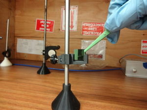
Fig. 3. Open-source sample holder v3
Fig. 4. Open-source fiber switcher
Fig. 5. Open-source rail
  • Shown on the left is an improved open-source sample holder for pieces of semiconductor wafers . Instead of the clamping a sample, it provides a 2mm lip that allows samples to fluently sink into the slot. The purpose of the design is to make sample switching faster and easier, and reduce abrasion caused by frequent sample switch. Also notice the hemisphere hole in the backside makes it easier for tweezers work. The design can be applied to variety of wafer-like material, we also use it as a mirror holder since it works fine with our appropediately sized mirror.
  • On the right is the improved fiber holder is a dual-channel fiber switcher, which attaches two fiber optic cable to a 8mm smooth rod and allow easy switching between cables for optical experiments.
  • In order to smoothly move each parts in our PL system, we've designed an optical rail that helps accurately align our system in a line and make lens focusing much easier. Installed with strong magnet the rail is clinging to the platform firmly. Now the system works in a fixed X-Y plane with measurable precision.

Further improvement

  • A component for a low temperature liquid to cool the sample during data taking.
  • The addition of a optical chopper and lock-in amplifier would improve signal to noise ratio
  • A monochromator would allow the system to focus on a small range of wavelengths at a time instead of looking a wide range of wavelengths.

As part of the open source optics project we will be designing all of these components.

To operate

See also

Cookies help us deliver our services. By using our services, you agree to our use of cookies.