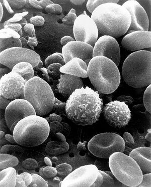
In the Material Physics Cleanroom (located in Stirling Hall atQueen's University), a Field Emission Scanning Electron Microscope is often used for imaging and characterization purposes. A scanning electron microscopeW uses an electron beam to obtain high levels of magnification and resolution for imaging. The specific type of FESEM found in the Materials Physics Cleanroom is the Leo/Zeiss 1530 Field Emission Scanning Electron Microscope[1] with Gemini gun and Nordliest EBSD.

Application notes:
- SEM works best with electrically conductive samples.
- SEM machines detect secondary electrons released from the interaction of the sample with the electron column beam
- Some SEM machines have an optional auger-electron detector; this one does not
- An EBSD detector (present on this machine) is used to read crystal structures at high energy
- SEM is a very qualitative measurement technique
- SEM is very fast at doing a scan, compared to AFM (atomic force microscope), with little required sample preparation
- This model of SEM is a field emission microscope; this allows for lower electron energies in the column, and therefore a wider range of samples can be scanned
- The SEM has two pumps: turbo pump for high pressure, and roughing pump for lower pressures. It can achieve 1e-6 torr vacuum, 1e-5 is also typical
- The energy of the electrons in this machine can be varried from 25KeV to 2KeV, which affects resolution of the image - a higher voltage will have a higher resolution
Microscope Specifications[edit | edit source]
- Schottky field-emission electron source
- Operating voltage: 0.2 - 30 kV
- Rated resolution: 1.2 nm at 20 kV, 2.5 nm at 5 kV, and 3 nm at 1 kV[2]
Protocol[edit | edit source]
- Working with the FESEM requires a trained lab researcher at all times.
- The lab researcher will assist in all of the imaging aspects and can offer helpful advice and guidance.
- The actual FESEM operation will be done by the researcher. A general outline of how the imaging program operates can be seen on this Georgia Tech College of Engineering page[3]
- Samples should be clean, prepared and free of debris before imaging. Some samples may require conductive coatings in order for the electron beam to properly image the subject.
- If possible, clean sample with ethanol to remove dust and oils.
- Always clean all instruments that will touch the sample with ethanol (ie. tweezers)
- A clean sample will pressurize very quickly.
- Always wear gloves if you are touching your samples: fingerprints make the vacuum chamber pressurize very slowly.
- A freshly cleaved edge should be used to obtain a cross-sectional scan. To cleave a sample, first use a diamond cutter to nick the sample. Use special flat cleaving tweezers to grip a large area of the sample on both sides of the nick. Roll the tweezers away from each other to cleave the sample along it's crystal plane. Do not touch the cleaved plane with anything. This cleaved edge should be tangential to the mounting plate, on the outer edge when mounted.
- Some movement of the image may be seen while scanning. This can be either the electron beam bending due to the properties of the sample, or the sample physically slipping around in the machine. Silver glue is best to obtain a secure mount.
Tips[edit | edit source]
- Be familiar with the general cleanroom procedures before entering. The dress attire conists of a cleanroom lab coat and booties.
- Always work with a qualified lab researcher unless proper training has been covered.
- If a cross sectional view is desired, ensure a clean break can be found (use a diamond scribe) on the sample and mount the sample in such an orientation that one edge slightly hangs off of the round holder.
- Double check sample identifications, sample placements and sample orientations before placing the samples inside the FESEM chamber.
Basic Operation Instructions[edit | edit source]
These instructions should only be followed by a trained operator, in the presence of a lab research.
The Software Screen[edit | edit source]
- Indicators at the bottom of the screen contain information and controls for vacuum levels, the electron column, among other things
- A bar contains information about the image: scale, magnification level, date, EHT voltage level (extra high tension, or high voltage) of the electron beam, WD (working distance, or the distance from the sample to the beam tip, also the focal distance of the beam)
- Buttons at the top control the gun and imaging. When a control is selected, the screen displays which mouse buttons control which option (LB = left mouse button, MB = middle scroll wheel button, RB = right mouse button). To change the option, hold down the associated mouse button and drag the mouse left or right.
- The first button is the magnification/focus controls.
- The second button is the contrast and brightness controls.
- The third button is stigmation which controls the ovality of the image, and is used as an advanced focussing technique
- Further to the right there are window controls buttons to make changes to the whole image (magnification, focus, etc) or just a small portion of it. Using the smaller portion allows faster rendering times.
The hardware controls[edit | edit source]
- Two joysticks control the movement of the sample within the chamber. It is labelled with which directions may move the sample closer to the beam. Note that the controls change when the sample is tilted for a cross-section scan.
- A mouse controls the other parts of the software.
SEM Basic Procedure[edit | edit source]
- Mount sample to be scanned on a mounting stub with either colloidal silver glue (permanent, takes a day to cure), or double-sided carbon tape (removable, instant).
- Attach the mounting stub to the mounting plate and gently screw it in place using the grub screw on the side of the mounting plate. Do not overtighten, as the vacuum will tighten the screw further.
- Pick up the mounting plate using the special mounting plate holder with the screw on the end. Screw it into the mounting plate.
- Open the door of the SEM.
- Slide the mounting plate into the platform in the SEM machine. This may require some wiggling and pushing.
- Close the chamber and hold it closed while the roughing pump starts, until it achieves vacuum. To start the pump use the software.
- Listen for the turbo pump to start as vacuum increases
- A green check mark will appear in the vacuum information slot on the indicators at the bottom of the software when full vacuum is achieved.
- Turn on the electron gun after the vacuum indicator has turned green
- Turn on EHT
- Scan your sample (see Scanning below)
- Move the sample away from the column
- Turn off EHT (the gun can stay on if you will be using the machine again soon)
- Turn off pump using the software.
- Listen to the (turbo) pump wind down - it takes approximately 1 minute, and starts very high pitched
- Release nitrogen into the vacuum chamber from the nitrogen tank. You should hear the gas entering the chamber.
- You will hear a poof sound when the vacuum has equalized enough to release the door seal. This can take a few minutes.
- Open the door and remove the mounting plate using the mounting plate holder tool; wiggle and pull it out of the grips
- Close the door.
- Remove your sample from the mounting stub.
- Clean up all tools and materials.
Scanning[edit | edit source]
- To scan your sample, first use the CCD camera view and joystick to orient the sample. The CCD camera is located at the back of the chamber
- If a different angle is desired, use the bars on the chamber door to manually rotate the mounting plate.
- Move the desired sample area into the column. Do not go too close to the column tip as hitting it would be a $20,000 repair - 10 cm is a good distance.
- Switch to the microscope view.
- To bring the image into focus, use the magnification, focus, contrast, and brightness controls. Try to focus initially on an interesting area of the sample to make it easier.
- Image colours are proportional to distance (nearer = brighter) and conductivity of different materials (move conductive = brighter)
- When a desired image is obtained, save it. The information bar will be stamped on the image.
Other Equipment in the Materials Physics Cleanroom[edit | edit source]
- X-Ray diffraction: Philips X'Pert XRD
- Atomic force microscopeW: Digital Instruments Multimode Nanoscope AFM
- Variable angle Spectroscopic EllipsometryW
For all booking information, rates and schedules, please see the Materials Physics cleanroom page.
Sources and Citations[edit | edit source]
- ↑ Material Physics Analytical Services. Material Physics Cleanroom, Queen's University. http://web.archive.org/web/20130421124904/http://www.physics.queensu.ca/%7erobbie/equipmentusage.html
- ↑ Characterization Equipment: Leo 1530 Field Emission Scanning Electron Microscope. National Nanotechnology Infrastructure Network at Penn State. http://web.archive.org/web/20100715150933/https://www.mri.psu.edu/facilities/NNIN/Equipment/EquipmentLEO1530FESEM.asp
- ↑ LEO 1530 Thermally-Assisted Field Emission (TFE) Scanning Electron Microscope (SEM). Materials Science and Engineering, Georgia Tech College of Engineering. http://web.archive.org/web/20100912170647/http://www.mse.gatech.edu:80/Research/Equipment_Facilities/CNC/LEO_1530/leo_1530.html